Gallery of new files
Jump to navigation
Jump to search
This special page shows the last uploaded files.
-
-
-
-
-
-
-
-
-
-
-
-
-
-

-
-
-

-

-

-

-

-

-
-

-

-

-

-

-
-
-
-

-

-
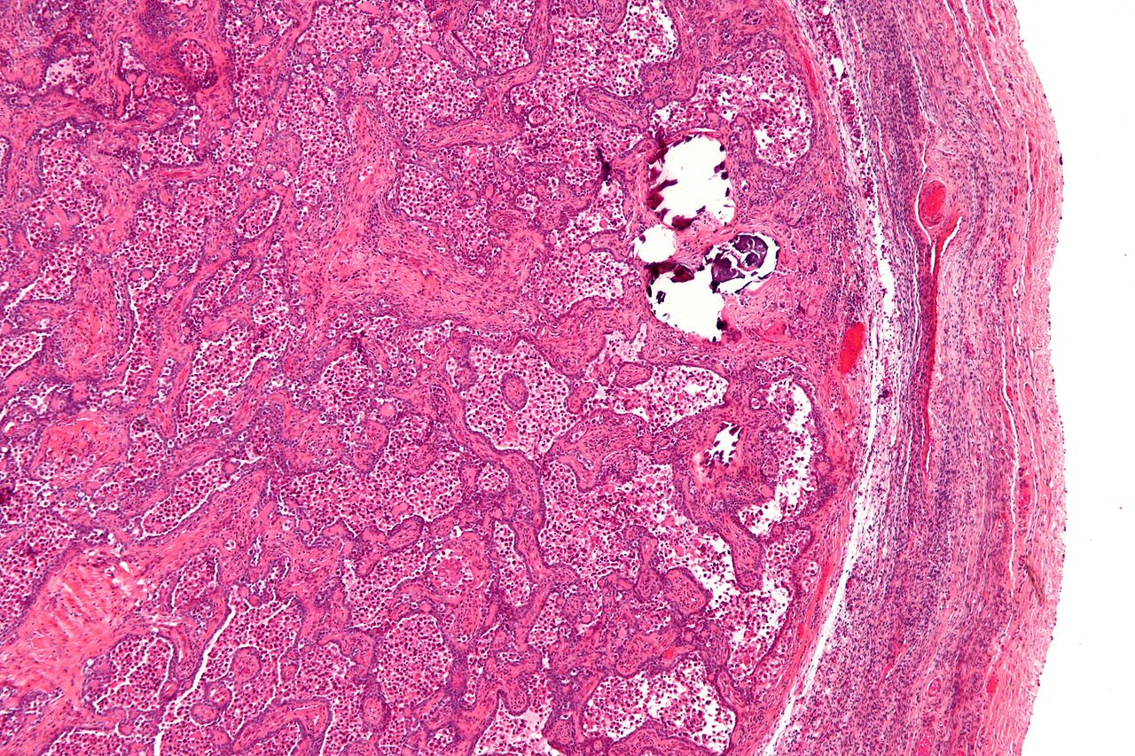
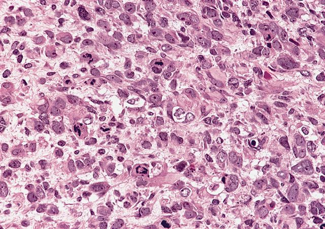
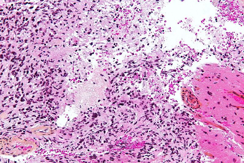
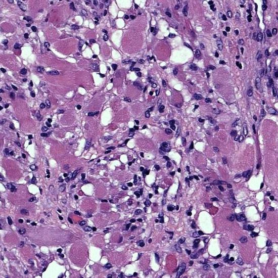
 1280px-Gonadoblastoma - high mag.jpg Sahar Memar Montazerin
15:28, 7 February 2019
1,280 × 853; 454 KB
-
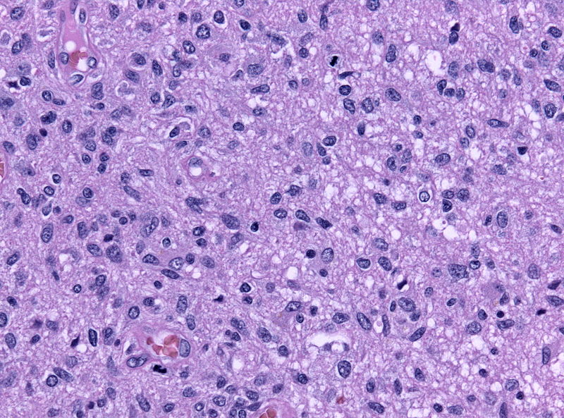
 1280px-Gonadoblastoma - low mag.jpg Sahar Memar Montazerin
15:24, 7 February 2019
1,280 × 853; 438 KB
-
-
-
-
-
-

-

-

-

-

-

-

-

-
-
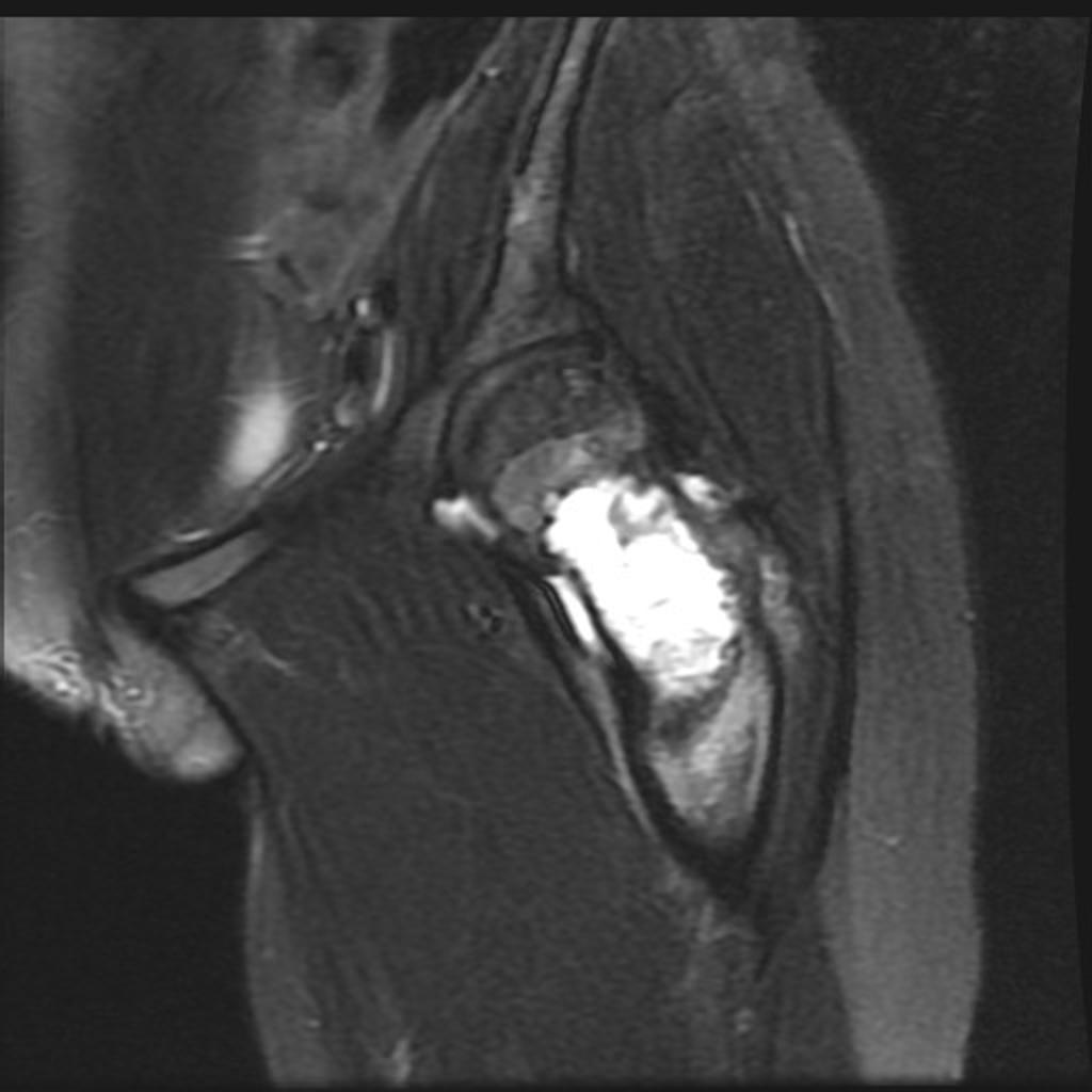
 Prostate-cancer-multiparametric-imaging-on-3-t-mri.jpg Farima Kahe
05:35, 6 February 2019
1,024 × 1,024; 86 KB
-

-
 Pleural and Pericardial Effusion CT.jpg Sabawoon Mirwais
23:37, 5 February 2019
1,024 × 1,024; 114 KB
-
-
-

-

-

-

-

-

-

-

-

-
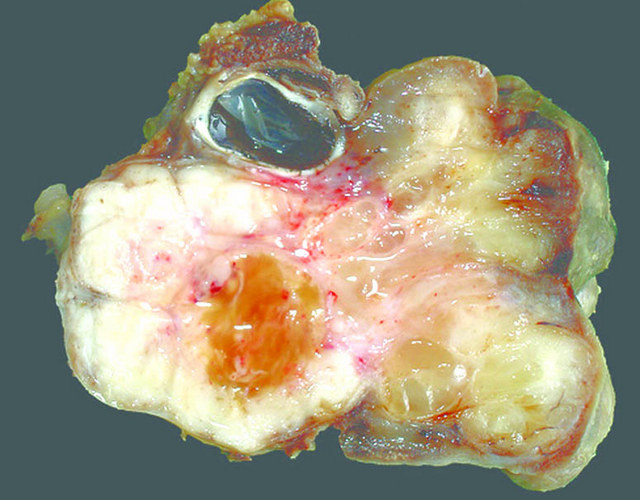
 Appendiceal-adenocarcinoma-complicated-by-retroperitoneal-abscess.jpg Soroush Seifirad
17:28, 5 February 2019
921 × 1,024; 183 KB
-

-
 Retro peritoneal abscess Complicated appendicitis.JPG Soroush Seifirad
17:04, 5 February 2019
986 × 921; 121 KB
-

-

-
-

-

-

-

-

-
-

-

-

-

-
-
-
-

-

-

-

-

-
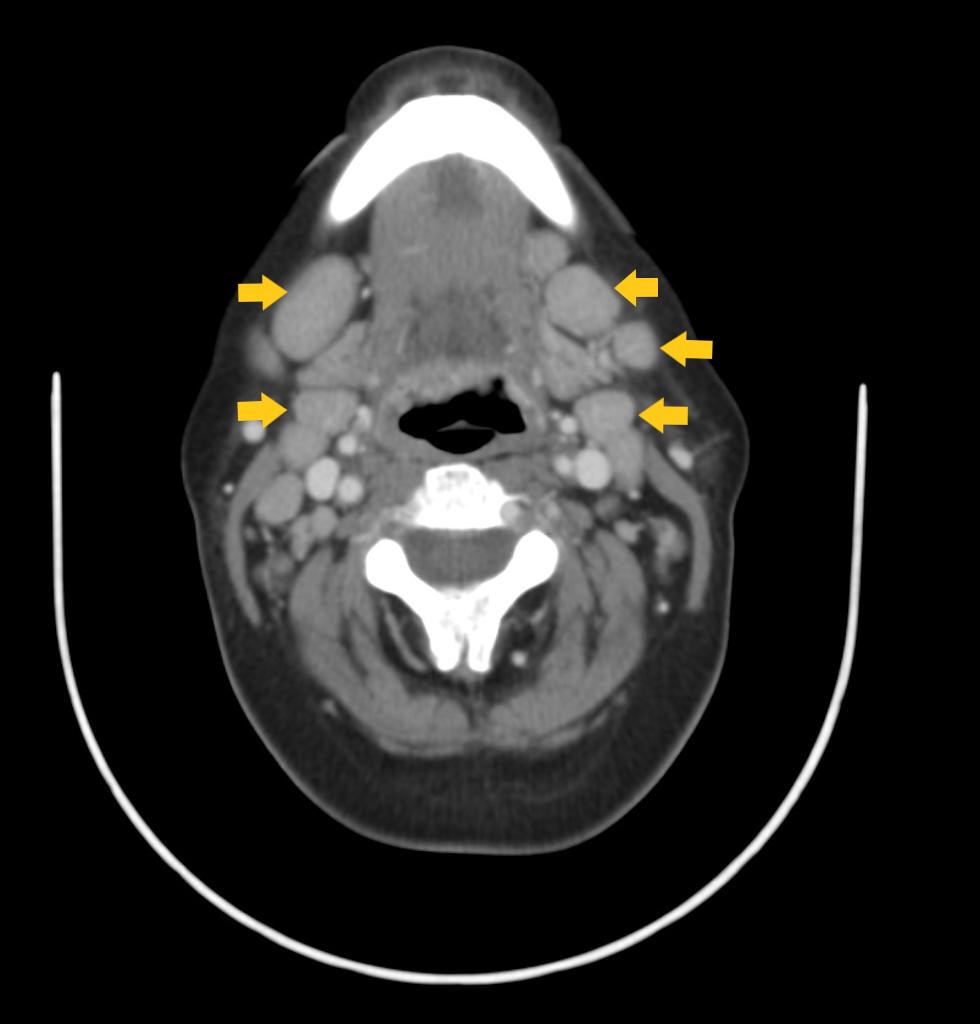
 Hypopharyngeal-squamous-cell-carcinoma CT.jpg Gertrude Djouka
17:48, 1 February 2019
1,024 × 1,024; 85 KB
-

-
-

-
 51075927 322309311724764 1632750230099197952 n.jpg Sara Mohsin
23:04, 31 January 2019
720 × 960; 123 KB
-
 51097708 344211846175715 6986947914031431680 n.jpg Sara Mohsin
21:19, 31 January 2019
1,080 × 1,340; 139 KB
-
-

-

-

-
-

-

-
-
 Acute-pancreatitis-and-walled-off-necrosis.jpg Sahar Memar Montazerin
16:59, 31 January 2019
1,024 × 747; 69 KB
-
-

-
 51261392 2194265964026264 868264017259397120 n.jpg Sara Mohsin
16:53, 31 January 2019
960 × 960; 83 KB
-
-

-

-

-

-
-
-
-
-

-
-

-
-

-

-

-
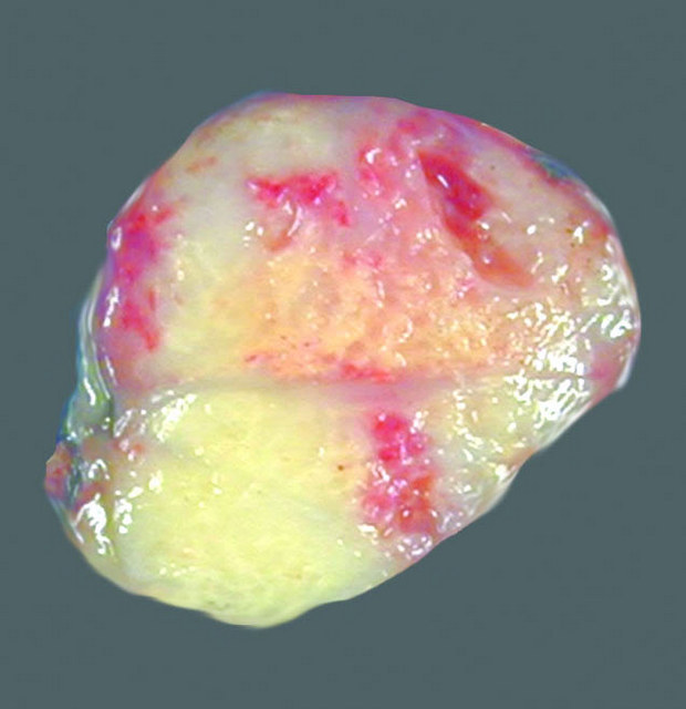
 Gross pathology of rhabdomyosarcoma.jpeg Shadan Mehraban
16:54, 30 January 2019
1,486 × 1,074; 310 KB
-
-

-

-
-
-
-
-
-

-

-

-

-

-

-
-
-

-
 Gross pathology of hypopharyngeal cancer.jpeg Gertrude Djouka
19:16, 24 January 2019
450 × 391; 19 KB
-
-

-

-
-
-
-
-

-
-
-
 Appendiceal-adenocarcinoma-complicated-by-retroperitoneal-abscess (4).jpg Soroush Seifirad
00:33, 23 January 2019
1,024 × 748; 205 KB
-
 Appendiceal-adenocarcinoma-complicated-by-retroperitoneal-abscess (2).jpg Soroush Seifirad
00:32, 23 January 2019
1,024 × 748; 183 KB
-
 Appendiceal-adenocarcinoma-complicated-by-retroperitoneal-abscess (1).jpg Soroush Seifirad
00:27, 23 January 2019
1,024 × 1,024; 253 KB
-
-
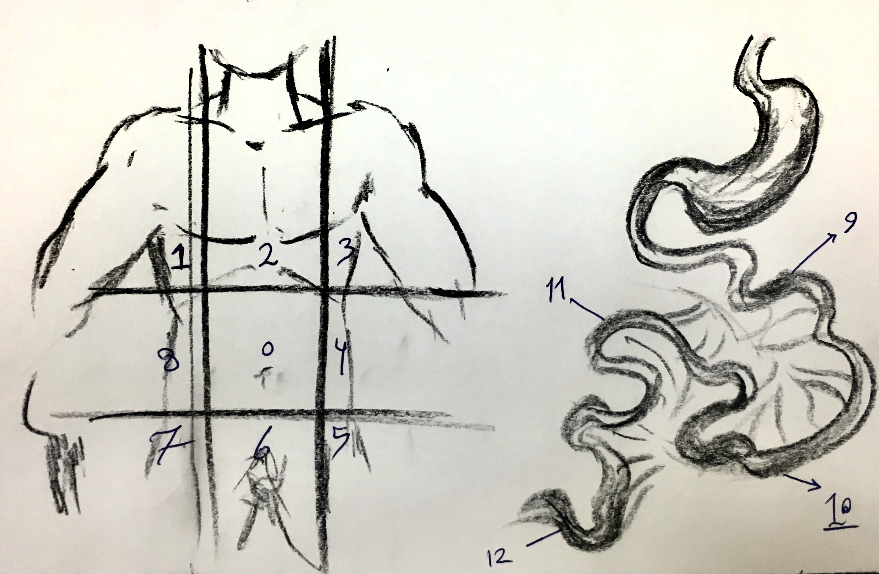 Peritoneal Carcinomatosis Index (PCI) Regions.jpg Soroush Seifirad
18:43, 22 January 2019
2,800 × 1,827; 1.21 MB
-
-

-

-
-

-

-
-

-
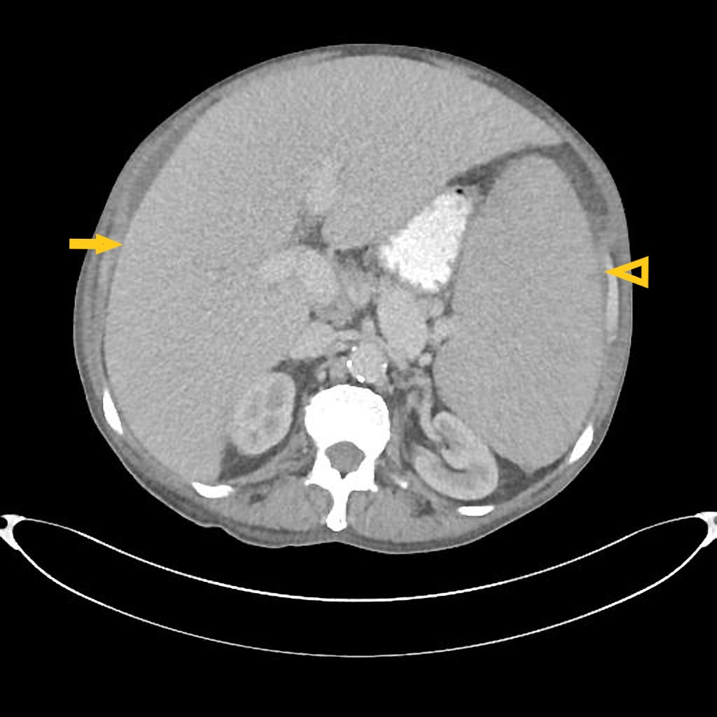
 CT of peritoneal carcinomatosis with peritoneal thickening and omental deposits.jpg Nima Nasiri
15:20, 21 January 2019
581 × 406; 62 KB
-
 Ovarian-cancer-with-peritoneal-carcinomatosis-1.jpg Nima Nasiri
15:11, 21 January 2019
1,024 × 1,024; 84 KB
-

-

-
-

-

-

-

-

-

-

-

-

-

-

-
-
-
-
-
-
-
-
-
-
-
-

-

-

-

-

-

-

-

-

-

-

-

-

-

-

-

-

-

-

-

-

-

-
-
 Diffuse large B-cell lymphoma of the small intestine (high mag).jpg Qurrat-ul-ain Abid
20:49, 14 January 2019
800 × 533; 175 KB
-
 Diffuse large B-cell lymphoma of the small intestine (intermed mag).jpg Qurrat-ul-ain Abid
20:42, 14 January 2019
800 × 533; 265 KB
-

-
-

-
 Gastrointestinal stromal tumor (GIST).jpg Qurrat-ul-ain Abid
15:12, 14 January 2019
800 × 533; 249 KB
-

-

-
-
-
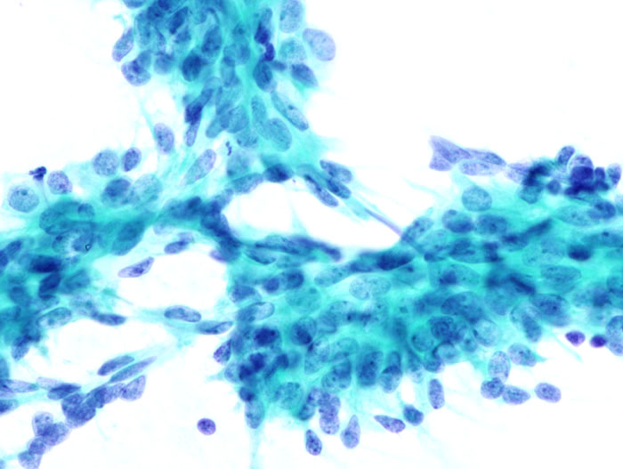
 Pleomorphic adenoma - cell block -- high mag.jpg Maneesha Nandimandalam
20:33, 10 January 2019
800 × 533; 167 KB
-

-

-
-

-

-

-

-

-

-

-

-

-

-

-

-
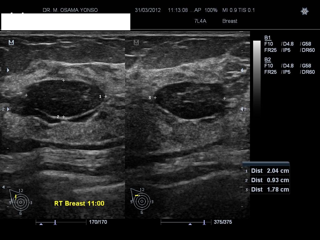
 Breast invasive lobular carcinoma ultrasound.gif Shadan Mehraban
16:29, 7 January 2019
960 × 720; 382 KB
-

-

-

-
-

-

-

-
-

-
-

-

-

-

-

-

-

-
 Microscopic feature of Sclerosing adenosis.jpg Shadan Mehraban
17:20, 3 January 2019
800 × 533; 180 KB
-

-
 Microscopic pathology of phyllodes tumor.jpg Shadan Mehraban
16:59, 3 January 2019
800 × 533; 179 KB
-

-
 Primary mediastinal large B-cell lymphoma CXR.PNG Badria Munir
02:21, 3 January 2019
960 × 720; 377 KB
-
-
 Bronchopneumonia caused by aspergillus.jpg Sindhuja palle
17:35, 2 January 2019
1,800 × 1,554; 267 KB
-
 Ct scan showing primary mediastinal large B-cell lymphoma.gif Badria Munir
17:42, 31 December 2018
1,280 × 720; 255 KB
-

-

-
-
 Primary mediastinal-lymphoma-large-b-cell-1.jpg Badria Munir
16:57, 31 December 2018
1,024 × 1,024; 88 KB
-

-
 Primary mediastinal large B-cell lymphoma .gif Badria Munir
16:44, 31 December 2018
1,280 × 720; 193 KB
-
-
-

-
-

-

-
 Multiple Carcinoid Tumors of the Small Bowel.jpg Qurrat-ul-ain Abid
17:18, 28 December 2018
550 × 203; 20 KB
-
 Small intestine neuroendocrine tumour.jpg Qurrat-ul-ain Abid
17:13, 28 December 2018
400 × 600; 119 KB
-

-
-
-
-
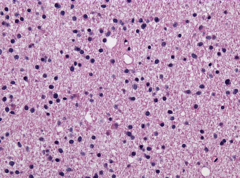
 800px-Pilocytic astrocytoma - smear - very high mag.jpg Fahimeh Shojaei
15:36, 28 December 2018
800 × 533; 147 KB
-
 Macroscopic-appearance-of-small-intestinal-adenocarcinoma-with-carcinomatosis-in-the.jpg Qurrat-ul-ain Abid
14:16, 28 December 2018
848 × 1,248; 129 KB
-
-
-
-
-
-

-

-
 Carcinoid Tumor of the Small Intestine.jpeg Qurrat-ul-ain Abid
14:19, 24 December 2018
299 × 209; 22 KB
-
-
-
-
-

-

-

-

-

-

-

-

-

-

-

-
 Fetal intermediate cellular type rhabdomyoma.jpg Nima Nasiri
16:47, 12 December 2018
550 × 550; 102 KB
-
-

-

-
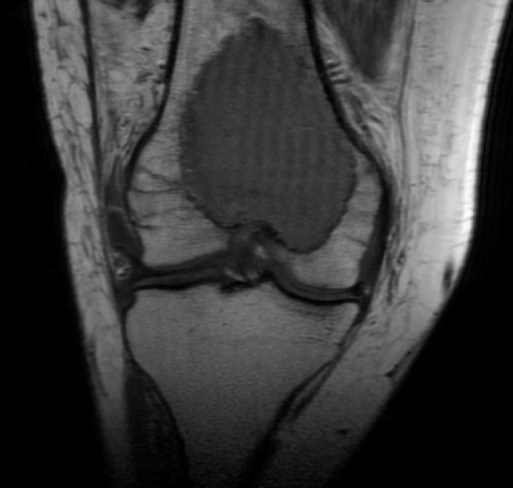
 MRI- non-displaced-distal-radial-fracture-.jpg Rohan Bhimani
21:55, 11 December 2018
1,024 × 1,024; 82 KB
-

-
 CT scan Lateral View with intraarticular step.JPG Rohan Bhimani
21:25, 11 December 2018
820 × 843; 98 KB
-

-
-
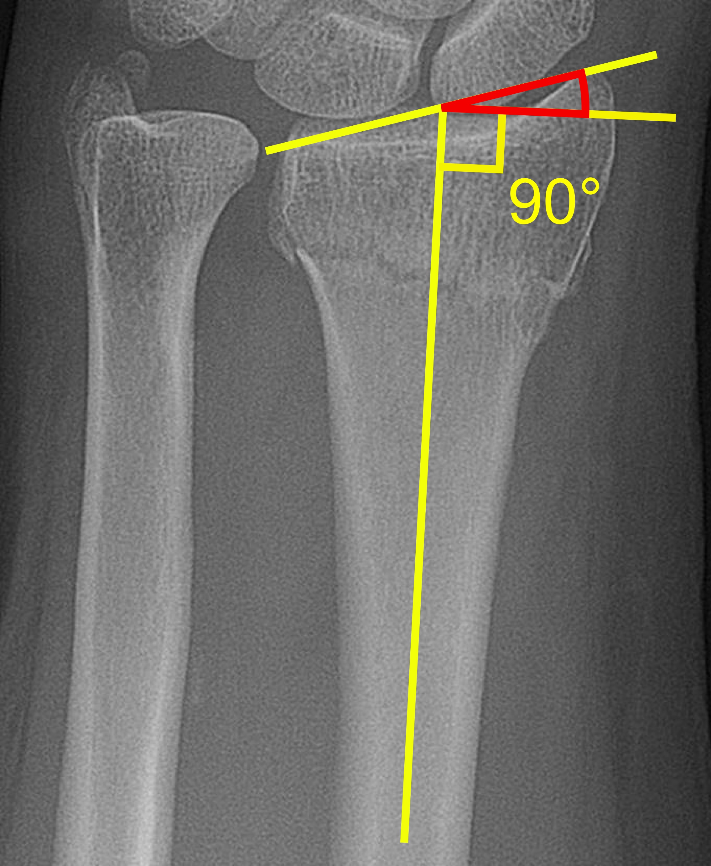 Radial inclination of distal radius fracture.jpg Rohan Bhimani
20:29, 11 December 2018
2,270 × 2,763; 1.07 MB
-
 Dorsal tilt of distal radius fracture.jpg Rohan Bhimani
20:23, 11 December 2018
2,070 × 4,283; 1.24 MB
-

-
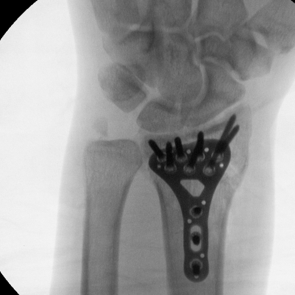
 Closed rduction of Distal radius fracture..png Rohan Bhimani
16:32, 11 December 2018
413 × 512; 175 KB
-
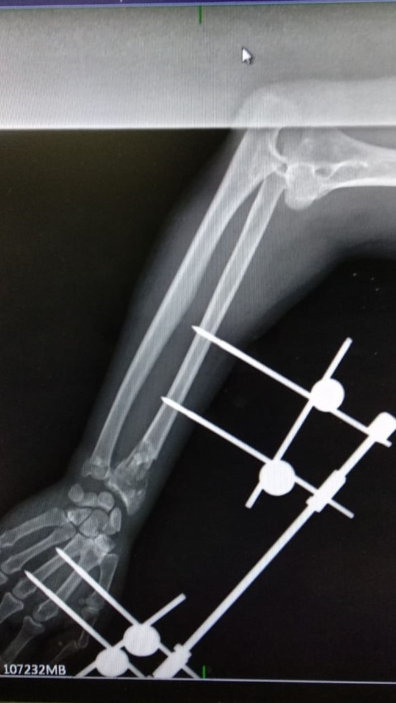 External fixation for Distsal end radius.jpg Rohan Bhimani
15:18, 11 December 2018
578 × 1,027; 111 KB
-

-
-

-
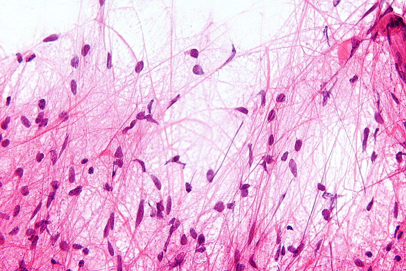
 Attaque; Periode Epileptoide. Planche XVII. Wellcome L0074938.jpg Fahimeh Shojaei
14:54, 11 December 2018
2,087 × 2,864; 1.05 MB
-

-

-

-

-

-
-

-

-

-

-
-

-
-
-
-

-

-
-
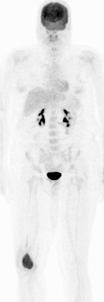
 PET scan -Thoracic endplate osteomyeltisdiscitis.jpg Rohan Bhimani
17:49, 6 December 2018
1,024 × 563; 45 KB
-
-
 MRI hemivertebra with congenital-scoliosis.jpg Rohan Bhimani
16:45, 6 December 2018
1,019 × 1,024; 126 KB
-
-
 CT scan Hemivertebra withcongenital-scoliosis.jpg Rohan Bhimani
15:17, 6 December 2018
1,024 × 974; 160 KB
-
-
 Hemivertebra-with-congenital-scoliosis.jpg Rohan Bhimani
21:44, 5 December 2018
1,024 × 1,024; 159 KB
-

-

-

-
 Transitional-cell-carcinoma-of-the-renal-pelvis.jpg Farima Kahe
15:48, 5 December 2018
1,024 × 1,014; 70 KB
-
-
-
-

-
-

-

-

-

-

-

-

-

-

-

-
-

-

-

-
-
-

-
-

-

-
 Archdischild-2004-September-89-9-809-F1.large.jpg Zahir Ali Shaikh
14:45, 13 November 2018
1,280 × 847; 112 KB
-
-

-
-
-

-

-

-

-

-

-

-
-
-

-

-

-

-
-

-

-

-
 Screenshot 2018-11-02 Myeloma - Cancer Stat Facts(1).png Hannan Javed
15:39, 2 November 2018
948 × 324; 19 KB
-
 Screenshot 2018-11-02 Myeloma - Cancer Stat Facts.png Hannan Javed
14:38, 2 November 2018
1,010 × 262; 24 KB
-
-
-
-

-
-

-

-
-

-
-

-

-
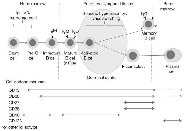 B lymphocytes differentaiation and maturation..jpg Hannan Javed
18:21, 23 October 2018
602 × 429; 28 KB
-

-

-
-

-

-

-
-
-

-
-
-
-
-
-
-
-
-
-
-
-

-
-

-

-
-

-

-
-

-

-

-
 Fibromuscular dysplasia microscopic pathology.jpg Mohsen Basiri
19:56, 24 August 2018
900 × 600; 647 KB
-

-
 Acute thrombotic microangiopathy - pas - high mag.jpg Sogand
14:16, 17 August 2018
1,200 × 800; 312 KB
-

-
-

-

-

-
-
-

-
-

-
-
-
-
-

-
-

-

-

-

-

-

-
-

-
-
-
-

-
-

-

-

-

-

-

-

-

-

-

-

-

-

-

-

-

-

-
-
-


.png)


.jpg)

.JPG)


.jpg)


.jpg)
.jpg)
.jpg)

_Regions.jpg)


.png)
.jpg)


.jpg)

.jpg)
.jpg)


.jpg)


.jpg)


.jpg)


.jpg)


.png)


.jpg)


.jpg)


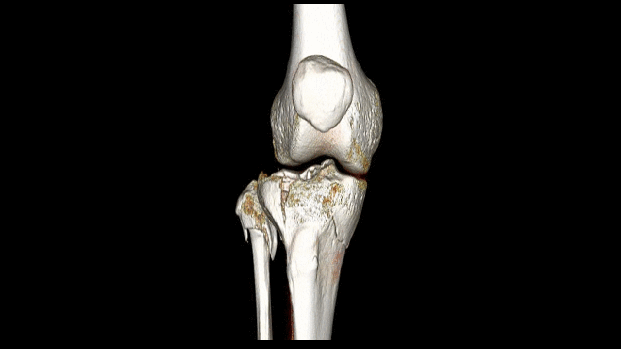
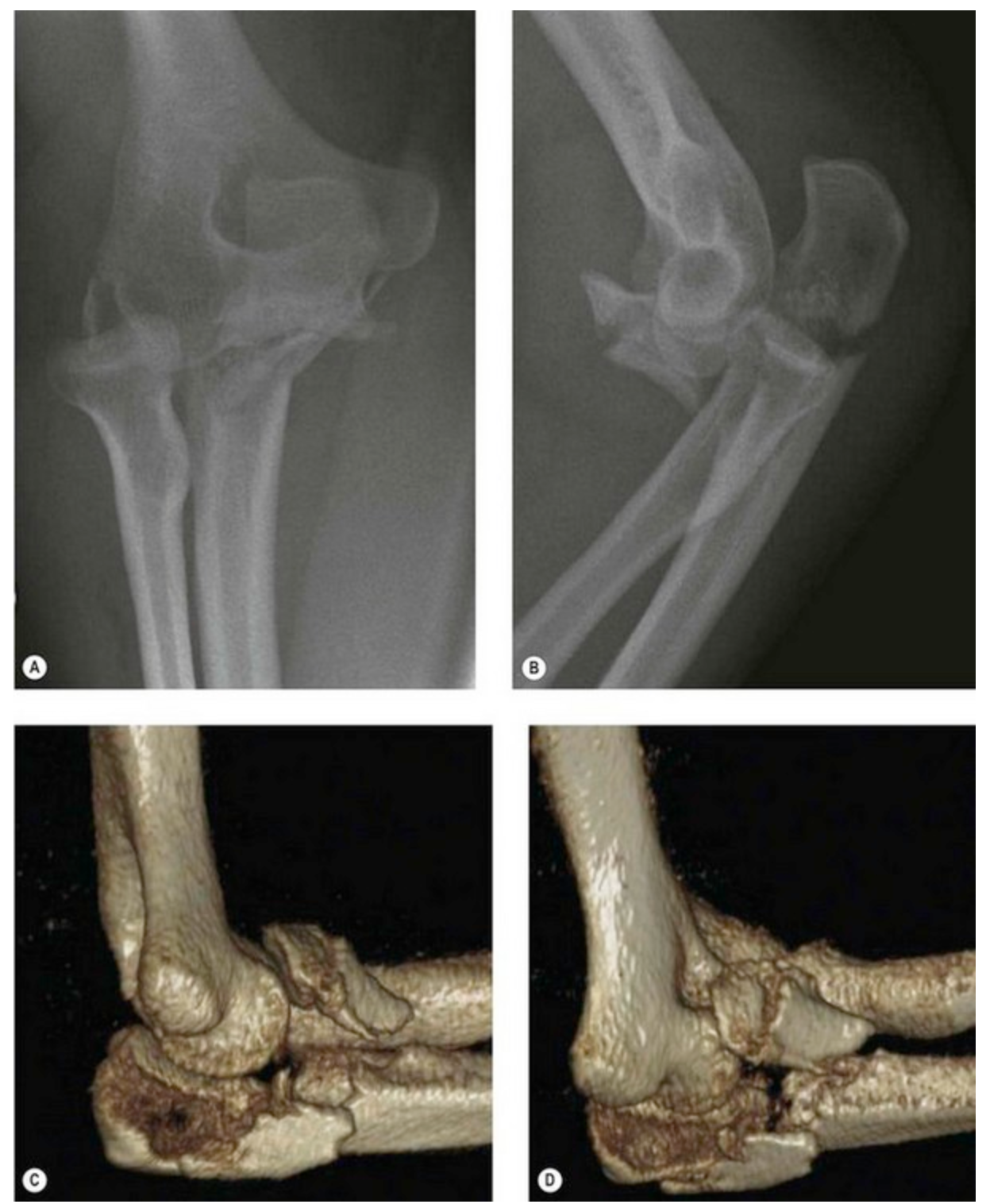
_The_radiographs_do_not_show_the_exact_anatomy_of_the_proximal_ulna_fracture._(C,_D)_CT_3-D_reconstruction_of_the_same_patient_clearly_identifies_the_fracture_morphology..png)
.png)