Rhabdomyosarcoma pathophysiology
|
Rhabdomyosarcoma Microchapters |
|
Diagnosis |
|---|
|
Treatment |
|
Case Studies |
|
Rhabdomyosarcoma pathophysiology On the Web |
|
American Roentgen Ray Society Images of Rhabdomyosarcoma pathophysiology |
|
Risk calculators and risk factors for Rhabdomyosarcoma pathophysiology |
Editor-In-Chief: C. Michael Gibson, M.S., M.D. [1];Associate Editor(s)-in-Chief: Suveenkrishna Pothuru, M.B,B.S. [2]
Overview
Rhabdomyosarcoma arises from the skeletal muscle cells. Development of alveolar rhabdomyosarcoma is result of specific genetic mutations. The pathogenesis of rhabdomyosarcoma include t(2;13) and t(1;13) chromosomal translocations. The microscopic pathology of rhabdomyosarcoma depends on the histological subtype.
Pathophysiology
Histology
- The origin of rhabdomyosarcoma is muscle cells.[1]
- The presentation sites of rhabdomyosarcoma are:
- Head and neck (28%)
- Extremities (24%)
- Genitourinary tract (18%)
- Trunk (11%)
- Orbit (7%)
- Retroperitoneum (6%)
- Other sites such as bladder, vagina, nasopharynx, and middle ear (3%)
Genetics
Alveolar rhabdomyosarcoma
Specific genetic abnormalities have been identified, that are specific for alveolar rhabdomyosarcomas. They include t(2;13) and t(1;13) chromosomal translocations resulting in PAX3-FKHR and PAX7-FKHR gene fusions.
Microscopic Pathology
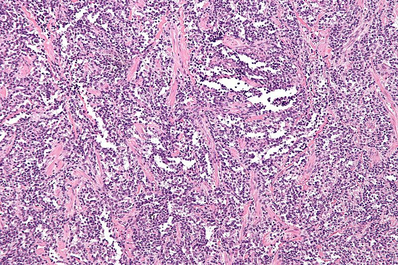
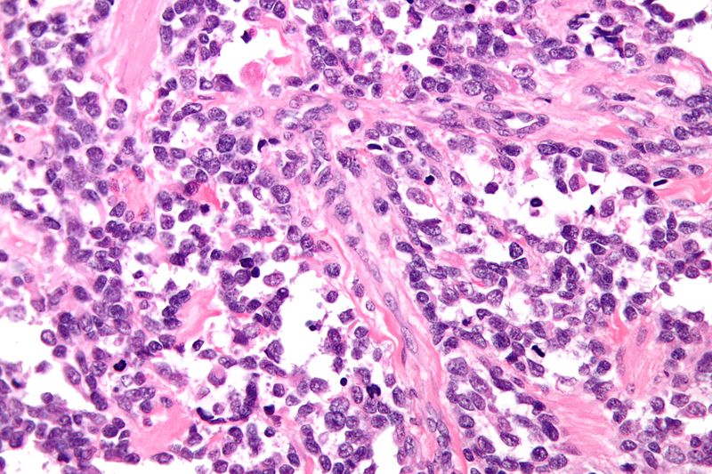
Characteristic features on microscopic analysis are variable depending on the rhabdomyosarcoma subtype:[2]
Alveolar Rhabdomyosarcoma
- Characterized by Alveolus-like pattern.
- Fibrous septae lined by tumor cells.
- Cells may "fall-off" the septa, i.e. be detached/scattered in the alveolus-like space.
- Space between fibrous septae may be filled with tumor, called solid variant of alveolar rhabdomyosarcoma.
- Rhabdomyoblasts: Essentially diagnostic cells with eccentric nucleus and moderate amount of intensely eosinophilic cytoplasm.
- Nuclear pleomorphism
- Mitoses
 |
 |
Embryonal Rhabdomyosarcoma
- Randomly arranged small cells
- Myxoid matrix
- Strap cells: Tadpole like morphology
- Rhabdomyoblasts: Essentially diagnostic cells with eccentric nucleus and moderate amount of intensely eosinophilic cytoplasm
Botryoid Rhabdomyosarcoma
- Malignant cells in an abundant myxoid stroma.
- Non-proliferating layer deep to the surface called "Cambium layer".
- Cambium layer is defined as cellular region deep to epithelial component.
Spindlecell Rhabdomyosarcoma
- Vesicular growth pattern
- Spindle cells
References
- ↑ Barr FG (1997). "Molecular genetics and pathogenesis of rhabdomyosarcoma". J Pediatr Hematol Oncol. 19 (6): 483–91. PMID 9407933.
- ↑ "librepathology".