Rhabdomyosarcoma pathophysiology: Difference between revisions
Ahmed Younes (talk | contribs) |
|||
| (12 intermediate revisions by one other user not shown) | |||
| Line 3: | Line 3: | ||
{{CMG}}; {{AE}} {{S.M}} | {{CMG}}; {{AE}} {{S.M}} | ||
==Overview== | ==Overview== | ||
The origin of [[rhabdomyosarcoma]] is straited [[muscle cells]].The presentation sites of rhabdomyosarcoma are [[head]] and [[neck]], [[extremities]], [[Genitourinary tract]], [[trunk]], [[orbit]], [[retroperitoneum]], [[bladder]], [[vagina]], [[nasopharynx]], and [[middle ear]].The exact [[pathogenesis]] of [[rhabdomyosarcoma]] is unclear. However, there is causal association between MET [[proto-oncogene]] and [[macrophage]] migration inhibitory factor (MIF) and it is thought that [[P53]] is related to [[oncogenic]] [[transformation]] and [[tumor]] progression. Multiple gene mutations are related to rhabdomyosarcoma such as [[loss of heterozygosity]] of 11p15, [[TP53]], [[NRAS]], [[KRAS]], [[HRAS]], PIK3CA, CTNNB1, FGFR4, and translocation in [[PAX3]] or [[PAX7]] genes with [[FOXO1]]. [[Immunohistochemistry]] can identify some specific proteins related to rhabdomyosarcoma such as [[desmin]], [[actin]], [[myogenin]], and [[myoglobin]]. The other [[histologic]] procedure is [[transmission electron microscopy]] (TEM) which can be used for poorly differentiated or undifferentiated [[tumors]] and myofilaments, actin, desmin, myotubular intermediate [[filaments]], and rudimentary Z-band material. [[Fusion protein]] is identified by [[Reverse transcription polymerase chain reaction|reverse transcriptase polymerase chain reaction]] and [[Fluorescence in situ hybridization|fluorescent in situ hybridization]]. | |||
==Pathophysiology== | ==Pathophysiology== | ||
| Line 9: | Line 9: | ||
===Histology=== | ===Histology=== | ||
* The origin of rhabdomyosarcoma is straited muscle cells.<ref name="pmid9407933">{{cite journal| author=Barr FG| title=Molecular genetics and pathogenesis of rhabdomyosarcoma. | journal=J Pediatr Hematol Oncol | year= 1997 | volume= 19 | issue= 6 | pages= 483-91 | pmid=9407933 | doi= | pmc= | url=https://www.ncbi.nlm.nih.gov/entrez/eutils/elink.fcgi?dbfrom=pubmed&tool=sumsearch.org/cite&retmode=ref&cmd=prlinks&id=9407933 }} </ref> | * The origin of [[rhabdomyosarcoma]] is straited [[muscle cells]].<ref name="pmid9407933">{{cite journal| author=Barr FG| title=Molecular genetics and pathogenesis of rhabdomyosarcoma. | journal=J Pediatr Hematol Oncol | year= 1997 | volume= 19 | issue= 6 | pages= 483-91 | pmid=9407933 | doi= | pmc= | url=https://www.ncbi.nlm.nih.gov/entrez/eutils/elink.fcgi?dbfrom=pubmed&tool=sumsearch.org/cite&retmode=ref&cmd=prlinks&id=9407933 }} </ref> | ||
* The presentation sites of rhabdomyosarcoma are: | * The presentation sites of rhabdomyosarcoma are: | ||
** Head and neck (28%) | ** [[Head]] and [[neck]] (28%) | ||
** Extremities (24%) | ** [[Extremities]] (24%) | ||
** Genitourinary tract (18%) | ** [[Genitourinary tract]] (18%) | ||
** Trunk (11%) | ** [[Trunk]] (11%) | ||
**Orbit (7%) | **[[Orbit]] (7%) | ||
**Retroperitoneum (6%) | **[[Retroperitoneum]] (6%) | ||
**Other sites such as bladder, vagina, nasopharynx, and middle ear (3%) | **Other sites such as [[bladder]], [[vagina]], [[nasopharynx]], and [[middle ear]] (3%) | ||
* Round or spindle-shaped muscle cells are present in embryonal rhabdomyosarcoma (ERMS).<ref name="pmid10666368">{{cite journal| author=Dias P, Chen B, Dilday B, Palmer H, Hosoi H, Singh S et al.| title=Strong immunostaining for myogenin in rhabdomyosarcoma is significantly associated with tumors of the alveolar subclass. | journal=Am J Pathol | year= 2000 | volume= 156 | issue= 2 | pages= 399-408 | pmid=10666368 | doi=10.1016/S0002-9440(10)64743-8 | pmc=1850049 | url=https://www.ncbi.nlm.nih.gov/entrez/eutils/elink.fcgi?dbfrom=pubmed&tool=sumsearch.org/cite&retmode=ref&cmd=prlinks&id=10666368 }} </ref> | * Round or spindle-shaped [[muscle cells]] are present in [[embryonal rhabdomyosarcoma]] (ERMS).<ref name="pmid10666368">{{cite journal| author=Dias P, Chen B, Dilday B, Palmer H, Hosoi H, Singh S et al.| title=Strong immunostaining for myogenin in rhabdomyosarcoma is significantly associated with tumors of the alveolar subclass. | journal=Am J Pathol | year= 2000 | volume= 156 | issue= 2 | pages= 399-408 | pmid=10666368 | doi=10.1016/S0002-9440(10)64743-8 | pmc=1850049 | url=https://www.ncbi.nlm.nih.gov/entrez/eutils/elink.fcgi?dbfrom=pubmed&tool=sumsearch.org/cite&retmode=ref&cmd=prlinks&id=10666368 }} </ref> | ||
* Alveolar architecture with small round undifferentiated cells which are | * [[Alveolar]] architecture with small round [[undifferentiated]] cells which are separated by dense hyalinized [[fibrous]] [[septa]] are present in [[alveolar rhabdomyosarcoma]] ([[ARMS]]). | ||
* Despite all muscle differentiation markers expression in ARMS, mature muscle characteristics is not seen in ARMS histology. | * Despite all muscle [[differentiation]] markers [[expression]] in [[ARMS]], mature muscle characteristics is not seen in [[ARMS]] [[histology]]. | ||
===Pathogenesis=== | ===Pathogenesis=== | ||
*The exact pathogenesis of rhabdomyosarcoma is unclear.<ref name="pmid16651427">{{cite journal| author=Taulli R, Scuoppo C, Bersani F, Accornero P, Forni PE, Miretti S et al.| title=Validation of met as a therapeutic target in alveolar and embryonal rhabdomyosarcoma. | journal=Cancer Res | year= 2006 | volume= 66 | issue= 9 | pages= 4742-9 | pmid=16651427 | doi=10.1158/0008-5472.CAN-05-4292 | pmc= | url=https://www.ncbi.nlm.nih.gov/entrez/eutils/elink.fcgi?dbfrom=pubmed&tool=sumsearch.org/cite&retmode=ref&cmd=prlinks&id=16651427 }} </ref> | *The exact [[pathogenesis]] of [[rhabdomyosarcoma]] is unclear.<ref name="pmid16651427">{{cite journal| author=Taulli R, Scuoppo C, Bersani F, Accornero P, Forni PE, Miretti S et al.| title=Validation of met as a therapeutic target in alveolar and embryonal rhabdomyosarcoma. | journal=Cancer Res | year= 2006 | volume= 66 | issue= 9 | pages= 4742-9 | pmid=16651427 | doi=10.1158/0008-5472.CAN-05-4292 | pmc= | url=https://www.ncbi.nlm.nih.gov/entrez/eutils/elink.fcgi?dbfrom=pubmed&tool=sumsearch.org/cite&retmode=ref&cmd=prlinks&id=16651427 }} </ref> | ||
*Distruption of skeletal muscle progenitor cell growth and related differentiation may cause rhabdomyosarcoma.<ref name="pmid16651427" /> | *Distruption of [[skeletal muscle]] progenitor cell growth and related differentiation may cause [[rhabdomyosarcoma]].<ref name="pmid16651427" /> | ||
*There is causal association between MET proto-oncogene and macrophage migration inhibitory factor (MIF).<ref name="pmid20861157">{{cite journal| author=Tarnowski M, Grymula K, Liu R, Tarnowska J, Drukala J, Ratajczak J et al.| title=Macrophage migration inhibitory factor is secreted by rhabdomyosarcoma cells, modulates tumor metastasis by binding to CXCR4 and CXCR7 receptors and inhibits recruitment of cancer-associated fibroblasts. | journal=Mol Cancer Res | year= 2010 | volume= 8 | issue= 10 | pages= 1328-43 | pmid=20861157 | doi=10.1158/1541-7786.MCR-10-0288 | pmc=2974061 | url=https://www.ncbi.nlm.nih.gov/entrez/eutils/elink.fcgi?dbfrom=pubmed&tool=sumsearch.org/cite&retmode=ref&cmd=prlinks&id=20861157 }} </ref> | *There is causal association between MET [[proto-oncogene]] and [[macrophage]] migration inhibitory factor (MIF).<ref name="pmid20861157">{{cite journal| author=Tarnowski M, Grymula K, Liu R, Tarnowska J, Drukala J, Ratajczak J et al.| title=Macrophage migration inhibitory factor is secreted by rhabdomyosarcoma cells, modulates tumor metastasis by binding to CXCR4 and CXCR7 receptors and inhibits recruitment of cancer-associated fibroblasts. | journal=Mol Cancer Res | year= 2010 | volume= 8 | issue= 10 | pages= 1328-43 | pmid=20861157 | doi=10.1158/1541-7786.MCR-10-0288 | pmc=2974061 | url=https://www.ncbi.nlm.nih.gov/entrez/eutils/elink.fcgi?dbfrom=pubmed&tool=sumsearch.org/cite&retmode=ref&cmd=prlinks&id=20861157 }} </ref> | ||
*It is thought that P53 is related to oncogenic transformation and tumor progression.<ref name="pmid20682800">{{cite journal| author=Xu J, Timares L, Heilpern C, Weng Z, Li C, Xu H et al.| title=Targeting wild-type and mutant p53 with small molecule CP-31398 blocks the growth of rhabdomyosarcoma by inducing reactive oxygen species-dependent apoptosis. | journal=Cancer Res | year= 2010 | volume= 70 | issue= 16 | pages= 6566-76 | pmid=20682800 | doi=10.1158/0008-5472.CAN-10-0942 | pmc=2922473 | url=https://www.ncbi.nlm.nih.gov/entrez/eutils/elink.fcgi?dbfrom=pubmed&tool=sumsearch.org/cite&retmode=ref&cmd=prlinks&id=20682800 }} </ref> | *It is thought that [[P53]] is related to [[oncogenic]] [[transformation]] and [[tumor]] progression.<ref name="pmid20682800">{{cite journal| author=Xu J, Timares L, Heilpern C, Weng Z, Li C, Xu H et al.| title=Targeting wild-type and mutant p53 with small molecule CP-31398 blocks the growth of rhabdomyosarcoma by inducing reactive oxygen species-dependent apoptosis. | journal=Cancer Res | year= 2010 | volume= 70 | issue= 16 | pages= 6566-76 | pmid=20682800 | doi=10.1158/0008-5472.CAN-10-0942 | pmc=2922473 | url=https://www.ncbi.nlm.nih.gov/entrez/eutils/elink.fcgi?dbfrom=pubmed&tool=sumsearch.org/cite&retmode=ref&cmd=prlinks&id=20682800 }} </ref> | ||
===Genetics=== | ===Genetics=== | ||
*According to microarray and targeted sequencing, following genomic characteristics are attributed to Rhabdomyosarcoma (RMS):<ref name="pmid2798419">{{cite journal| author=Scrable H, Cavenee W, Ghavimi F, Lovell M, Morgan K, Sapienza C| title=A model for embryonal rhabdomyosarcoma tumorigenesis that involves genome imprinting. | journal=Proc Natl Acad Sci U S A | year= 1989 | volume= 86 | issue= 19 | pages= 7480-4 | pmid=2798419 | doi= | pmc=298088 | url=https://www.ncbi.nlm.nih.gov/entrez/eutils/elink.fcgi?dbfrom=pubmed&tool=sumsearch.org/cite&retmode=ref&cmd=prlinks&id=2798419 }} </ref><ref name="pmid10918230">{{cite journal| author=Taylor AC, Shu L, Danks MK, Poquette CA, Shetty S, Thayer MJ et al.| title=P53 mutation and MDM2 amplification frequency in pediatric rhabdomyosarcoma tumors and cell lines. | journal=Med Pediatr Oncol | year= 2000 | volume= 35 | issue= 2 | pages= 96-103 | pmid=10918230 | doi= | pmc= | url=https://www.ncbi.nlm.nih.gov/entrez/eutils/elink.fcgi?dbfrom=pubmed&tool=sumsearch.org/cite&retmode=ref&cmd=prlinks&id=10918230 }} </ref><ref name="pmid2680062">{{cite journal| author=Stratton MR, Fisher C, Gusterson BA, Cooper CS| title=Detection of point mutations in N-ras and K-ras genes of human embryonal rhabdomyosarcomas using oligonucleotide probes and the polymerase chain reaction. | journal=Cancer Res | year= 1989 | volume= 49 | issue= 22 | pages= 6324-7 | pmid=2680062 | doi= | pmc= | url=https://www.ncbi.nlm.nih.gov/entrez/eutils/elink.fcgi?dbfrom=pubmed&tool=sumsearch.org/cite&retmode=ref&cmd=prlinks&id=2680062 }} </ref><ref name="pmid22142829">{{cite journal| author=Shukla N, Ameur N, Yilmaz I, Nafa K, Lau CY, Marchetti A et al.| title=Oncogene mutation profiling of pediatric solid tumors reveals significant subsets of embryonal rhabdomyosarcoma and neuroblastoma with mutated genes in growth signaling pathways. | journal=Clin Cancer Res | year= 2012 | volume= 18 | issue= 3 | pages= 748-57 | pmid=22142829 | doi=10.1158/1078-0432.CCR-11-2056 | pmc=3271129 | url=https://www.ncbi.nlm.nih.gov/entrez/eutils/elink.fcgi?dbfrom=pubmed&tool=sumsearch.org/cite&retmode=ref&cmd=prlinks&id=22142829 }} </ref><ref name="pmid19809159">{{cite journal| author=Taylor JG, Cheuk AT, Tsang PS, Chung JY, Song YK, Desai K et al.| title=Identification of FGFR4-activating mutations in human rhabdomyosarcomas that promote metastasis in xenotransplanted models. | journal=J Clin Invest | year= 2009 | volume= 119 | issue= 11 | pages= 3395-407 | pmid=19809159 | doi=10.1172/JCI39703 | pmc=2769177 | url=https://www.ncbi.nlm.nih.gov/entrez/eutils/elink.fcgi?dbfrom=pubmed&tool=sumsearch.org/cite&retmode=ref&cmd=prlinks&id=19809159 }} </ref><ref name="pmid8098985">{{cite journal| author=Barr FG, Galili N, Holick J, Biegel JA, Rovera G, Emanuel BS| title=Rearrangement of the PAX3 paired box gene in the paediatric solid tumour alveolar rhabdomyosarcoma. | journal=Nat Genet | year= 1993 | volume= 3 | issue= 2 | pages= 113-7 | pmid=8098985 | doi=10.1038/ng0293-113 | pmc= | url=https://www.ncbi.nlm.nih.gov/entrez/eutils/elink.fcgi?dbfrom=pubmed&tool=sumsearch.org/cite&retmode=ref&cmd=prlinks&id=8098985 }} </ref> | *According to [[microarray]] and targeted [[sequencing]], following [[genomic]] characteristics are attributed to [[Rhabdomyosarcoma]] (RMS):<ref name="pmid2798419">{{cite journal| author=Scrable H, Cavenee W, Ghavimi F, Lovell M, Morgan K, Sapienza C| title=A model for embryonal rhabdomyosarcoma tumorigenesis that involves genome imprinting. | journal=Proc Natl Acad Sci U S A | year= 1989 | volume= 86 | issue= 19 | pages= 7480-4 | pmid=2798419 | doi= | pmc=298088 | url=https://www.ncbi.nlm.nih.gov/entrez/eutils/elink.fcgi?dbfrom=pubmed&tool=sumsearch.org/cite&retmode=ref&cmd=prlinks&id=2798419 }} </ref><ref name="pmid10918230">{{cite journal| author=Taylor AC, Shu L, Danks MK, Poquette CA, Shetty S, Thayer MJ et al.| title=P53 mutation and MDM2 amplification frequency in pediatric rhabdomyosarcoma tumors and cell lines. | journal=Med Pediatr Oncol | year= 2000 | volume= 35 | issue= 2 | pages= 96-103 | pmid=10918230 | doi= | pmc= | url=https://www.ncbi.nlm.nih.gov/entrez/eutils/elink.fcgi?dbfrom=pubmed&tool=sumsearch.org/cite&retmode=ref&cmd=prlinks&id=10918230 }} </ref><ref name="pmid2680062">{{cite journal| author=Stratton MR, Fisher C, Gusterson BA, Cooper CS| title=Detection of point mutations in N-ras and K-ras genes of human embryonal rhabdomyosarcomas using oligonucleotide probes and the polymerase chain reaction. | journal=Cancer Res | year= 1989 | volume= 49 | issue= 22 | pages= 6324-7 | pmid=2680062 | doi= | pmc= | url=https://www.ncbi.nlm.nih.gov/entrez/eutils/elink.fcgi?dbfrom=pubmed&tool=sumsearch.org/cite&retmode=ref&cmd=prlinks&id=2680062 }} </ref><ref name="pmid22142829">{{cite journal| author=Shukla N, Ameur N, Yilmaz I, Nafa K, Lau CY, Marchetti A et al.| title=Oncogene mutation profiling of pediatric solid tumors reveals significant subsets of embryonal rhabdomyosarcoma and neuroblastoma with mutated genes in growth signaling pathways. | journal=Clin Cancer Res | year= 2012 | volume= 18 | issue= 3 | pages= 748-57 | pmid=22142829 | doi=10.1158/1078-0432.CCR-11-2056 | pmc=3271129 | url=https://www.ncbi.nlm.nih.gov/entrez/eutils/elink.fcgi?dbfrom=pubmed&tool=sumsearch.org/cite&retmode=ref&cmd=prlinks&id=22142829 }} </ref><ref name="pmid19809159">{{cite journal| author=Taylor JG, Cheuk AT, Tsang PS, Chung JY, Song YK, Desai K et al.| title=Identification of FGFR4-activating mutations in human rhabdomyosarcomas that promote metastasis in xenotransplanted models. | journal=J Clin Invest | year= 2009 | volume= 119 | issue= 11 | pages= 3395-407 | pmid=19809159 | doi=10.1172/JCI39703 | pmc=2769177 | url=https://www.ncbi.nlm.nih.gov/entrez/eutils/elink.fcgi?dbfrom=pubmed&tool=sumsearch.org/cite&retmode=ref&cmd=prlinks&id=19809159 }} </ref><ref name="pmid8098985">{{cite journal| author=Barr FG, Galili N, Holick J, Biegel JA, Rovera G, Emanuel BS| title=Rearrangement of the PAX3 paired box gene in the paediatric solid tumour alveolar rhabdomyosarcoma. | journal=Nat Genet | year= 1993 | volume= 3 | issue= 2 | pages= 113-7 | pmid=8098985 | doi=10.1038/ng0293-113 | pmc= | url=https://www.ncbi.nlm.nih.gov/entrez/eutils/elink.fcgi?dbfrom=pubmed&tool=sumsearch.org/cite&retmode=ref&cmd=prlinks&id=8098985 }} </ref> | ||
**Loss of heterozygosity of 11p15. | **[[Loss of heterozygosity]] of 11p15. | ||
** Mutations in TP53 | ** [[Mutations]] in [[TP53]] | ||
** Mutations in NRAS | ** [[Mutations]] in [[NRAS]] | ||
** Mutations in KRAS | ** [[Mutations]] in [[KRAS]] | ||
** Mutations in HRAS | ** [[Mutations]] in [[HRAS]] | ||
** Mutations in PIK3CA | ** [[Mutations]] in PIK3CA | ||
** Mutations in CTNNB1 | ** [[Mutations]] in CTNNB1 | ||
** Mutations in FGFR4 | ** [[Mutations]] in FGFR4 | ||
** Translocations in PAX3 or PAX7 genes with FOXO1 | ** [[Translocations]] in [[PAX3]] or [[PAX7]] genes with [[FOXO1]] | ||
*Specific genetic changes in different types of rhabdomyosarcoma: | *Specific genetic changes in different types of [[rhabdomyosarcoma]]: | ||
*Alveolar rhabdomyosarcoma:<ref name="pmid22454413">{{cite journal| author=Missiaglia E, Williamson D, Chisholm J, Wirapati P, Pierron G, Petel F et al.| title=PAX3/FOXO1 fusion gene status is the key prognostic molecular marker in rhabdomyosarcoma and significantly improves current risk stratification. | journal=J Clin Oncol | year= 2012 | volume= 30 | issue= 14 | pages= 1670-7 | pmid=22454413 | doi=10.1200/JCO.2011.38.5591 | pmc= | url=https://www.ncbi.nlm.nih.gov/entrez/eutils/elink.fcgi?dbfrom=pubmed&tool=sumsearch.org/cite&retmode=ref&cmd=prlinks&id=22454413 }} </ref><ref name="pmid20663909">{{cite journal| author=Cao L, Yu Y, Bilke S, Walker RL, Mayeenuddin LH, Azorsa DO et al.| title=Genome-wide identification of PAX3-FKHR binding sites in rhabdomyosarcoma reveals candidate target genes important for development and cancer. | journal=Cancer Res | year= 2010 | volume= 70 | issue= 16 | pages= 6497-508 | pmid=20663909 | doi=10.1158/0008-5472.CAN-10-0582 | pmc=2922412 | url=https://www.ncbi.nlm.nih.gov/entrez/eutils/elink.fcgi?dbfrom=pubmed&tool=sumsearch.org/cite&retmode=ref&cmd=prlinks&id=20663909 }} </ref> | *[[Alveolar rhabdomyosarcoma]]:<ref name="pmid22454413">{{cite journal| author=Missiaglia E, Williamson D, Chisholm J, Wirapati P, Pierron G, Petel F et al.| title=PAX3/FOXO1 fusion gene status is the key prognostic molecular marker in rhabdomyosarcoma and significantly improves current risk stratification. | journal=J Clin Oncol | year= 2012 | volume= 30 | issue= 14 | pages= 1670-7 | pmid=22454413 | doi=10.1200/JCO.2011.38.5591 | pmc= | url=https://www.ncbi.nlm.nih.gov/entrez/eutils/elink.fcgi?dbfrom=pubmed&tool=sumsearch.org/cite&retmode=ref&cmd=prlinks&id=22454413 }} </ref><ref name="pmid20663909">{{cite journal| author=Cao L, Yu Y, Bilke S, Walker RL, Mayeenuddin LH, Azorsa DO et al.| title=Genome-wide identification of PAX3-FKHR binding sites in rhabdomyosarcoma reveals candidate target genes important for development and cancer. | journal=Cancer Res | year= 2010 | volume= 70 | issue= 16 | pages= 6497-508 | pmid=20663909 | doi=10.1158/0008-5472.CAN-10-0582 | pmc=2922412 | url=https://www.ncbi.nlm.nih.gov/entrez/eutils/elink.fcgi?dbfrom=pubmed&tool=sumsearch.org/cite&retmode=ref&cmd=prlinks&id=20663909 }} </ref> | ||
**Translocation of FKHR gene on chromosome 13 with the PAX3 gene on chromosome 2 or the PAX7 gene on chromosome 1 | **[[Translocation]] of FKHR [[gene]] on [[Chromosome 13 (human)|chromosome 13]] with the [[PAX3]] [[gene]] on [[chromosome 2]] or the [[PAX7]] [[gene]] on [[chromosome 1]] | ||
**PAX7 translocation occur in younger patients with longer event-free survival rate | **[[PAX7]] [[Translocations|translocation]] occur in younger patients with longer event-free [[survival rate]] | ||
*Embryonal rhabdomyosarcoma:<ref name="pmid94079332" /> | *[[Embryonal rhabdomyosarcoma]]:<ref name="pmid94079332" /> | ||
** Particular gene changes are not clear | ** Particular [[gene]] changes are not clear | ||
** | **[[Loss of heterozygosity]] from the 11p15 region | ||
** Hyperdiploid gene | ** Hyperdiploid [[gene]] | ||
** Lack of gene amplification | ** Lack of [[gene]] [[amplification]] | ||
==Associated conditions== | ==Associated conditions== | ||
===Immunohistochemistry=== | ===Immunohistochemistry=== | ||
* Through immunohistochemistry, muscle specific proteins can be identified in RMS.<ref name="pmid10666368">{{cite journal| author=Dias P, Chen B, Dilday B, Palmer H, Hosoi H, Singh S et al.| title=Strong immunostaining for myogenin in rhabdomyosarcoma is significantly associated with tumors of the alveolar subclass. | journal=Am J Pathol | year= 2000 | volume= 156 | issue= 2 | pages= 399-408 | pmid=10666368 | doi=10.1016/S0002-9440(10)64743-8 | pmc=1850049 | url=https://www.ncbi.nlm.nih.gov/entrez/eutils/elink.fcgi?dbfrom=pubmed&tool=sumsearch.org/cite&retmode=ref&cmd=prlinks&id=10666368 }} </ref> | * Through [[immunohistochemistry]], muscle specific proteins can be identified in RMS.<ref name="pmid10666368">{{cite journal| author=Dias P, Chen B, Dilday B, Palmer H, Hosoi H, Singh S et al.| title=Strong immunostaining for myogenin in rhabdomyosarcoma is significantly associated with tumors of the alveolar subclass. | journal=Am J Pathol | year= 2000 | volume= 156 | issue= 2 | pages= 399-408 | pmid=10666368 | doi=10.1016/S0002-9440(10)64743-8 | pmc=1850049 | url=https://www.ncbi.nlm.nih.gov/entrez/eutils/elink.fcgi?dbfrom=pubmed&tool=sumsearch.org/cite&retmode=ref&cmd=prlinks&id=10666368 }} </ref> | ||
* RMS stains positive for following proteins: | * RMS stains positive for following [[proteins]]: | ||
** | ** [[Polyclonal]] [[desmin]] (99%) | ||
** Muscle-specific actin (95%) | ** Muscle-specific [[actin]] (95%) | ||
** Myogenin (95%) | ** [[Myogenin]] or [[myogenic]] differentiation 1 ( MyoD1) (95%) | ||
*** Expressed more in ARMS rather than ERMS | *** Expressed more in [[ARMS]] rather than ERMS | ||
*** Associated with poorer prognosis | *** Associated with poorer [[prognosis]] | ||
** Myoglobin (78%) | ** [[Myoglobin]] (78%) | ||
===Transmission electron microscopy=== | ===Transmission electron microscopy=== | ||
* Transmission electron microscopy (TEM) can be used for poorly differentiated or undifferentiated tumors.<ref name="pmid12110339">{{cite journal| author=Hicks J, Flaitz C| title=Rhabdomyosarcoma of the head and neck in children. | journal=Oral Oncol | year= 2002 | volume= 38 | issue= 5 | pages= 450-9 | pmid=12110339 | doi= | pmc= | url=https://www.ncbi.nlm.nih.gov/entrez/eutils/elink.fcgi?dbfrom=pubmed&tool=sumsearch.org/cite&retmode=ref&cmd=prlinks&id=12110339 }} </ref><ref name="pmid11353062">{{cite journal| author=Parham DM| title=Pathologic classification of rhabdomyosarcomas and correlations with molecular studies. | journal=Mod Pathol | year= 2001 | volume= 14 | issue= 5 | pages= 506-14 | pmid=11353062 | doi=10.1038/modpathol.3880339 | pmc= | url=https://www.ncbi.nlm.nih.gov/entrez/eutils/elink.fcgi?dbfrom=pubmed&tool=sumsearch.org/cite&retmode=ref&cmd=prlinks&id=11353062 }} </ref> | * [[Transmission electron microscopy]] (TEM) can be used for poorly differentiated or undifferentiated [[tumors]].<ref name="pmid12110339">{{cite journal| author=Hicks J, Flaitz C| title=Rhabdomyosarcoma of the head and neck in children. | journal=Oral Oncol | year= 2002 | volume= 38 | issue= 5 | pages= 450-9 | pmid=12110339 | doi= | pmc= | url=https://www.ncbi.nlm.nih.gov/entrez/eutils/elink.fcgi?dbfrom=pubmed&tool=sumsearch.org/cite&retmode=ref&cmd=prlinks&id=12110339 }} </ref><ref name="pmid11353062">{{cite journal| author=Parham DM| title=Pathologic classification of rhabdomyosarcomas and correlations with molecular studies. | journal=Mod Pathol | year= 2001 | volume= 14 | issue= 5 | pages= 506-14 | pmid=11353062 | doi=10.1038/modpathol.3880339 | pmc= | url=https://www.ncbi.nlm.nih.gov/entrez/eutils/elink.fcgi?dbfrom=pubmed&tool=sumsearch.org/cite&retmode=ref&cmd=prlinks&id=11353062 }} </ref> | ||
* | * Presence of following proteins are associated with [[TEM]] features of RMS: | ||
** Myofilaments | ** Myofilaments | ||
** Desmin and actin filaments | ** [[Desmin]] and [[actin]] [[filaments]] | ||
** Myotubular intermediate filaments | ** Myotubular intermediate [[filaments]] | ||
** Rudimentary Z-band material | ** Rudimentary Z-band material | ||
===Reverse transcriptase polymerase chain reaction (RT-PCR)=== | ===Reverse transcriptase polymerase chain reaction (RT-PCR)=== | ||
===Fluorescent in situ hybridization (FISH) === | ===Fluorescent in situ hybridization (FISH) === | ||
*By using RT-PCR and FISH, features of fusion | *By using [[RT-PCR]] and [[FISH]], features of [[fusion protein]] are identified in [[ARMS]].<ref name="pmid12951587">{{cite journal| author=Helman LJ, Meltzer P| title=Mechanisms of sarcoma development. | journal=Nat Rev Cancer | year= 2003 | volume= 3 | issue= 9 | pages= 685-94 | pmid=12951587 | doi=10.1038/nrc1168 | pmc= | url=https://www.ncbi.nlm.nih.gov/entrez/eutils/elink.fcgi?dbfrom=pubmed&tool=sumsearch.org/cite&retmode=ref&cmd=prlinks&id=12951587 }} </ref> | ||
==Gross Pathology== | ==Gross Pathology== | ||
{| class="wikitable" | |||
|+ | |||
! style="background:#4479BA; color: #FFFFFF;" align="center" + |Gross findings | |||
[[File:Embryonal rhabdomyosarcoma.jpg|thumb|none|300px| Gross feature of orbital embryonal rhabdomyosarcoma [http://www.pathologyoutlines.com/topic/softtissueembryonalrhabdo.html Source: Contributed by Mark R. Wick, M.D., from Pathologyoutlines]]] | ! style="background:#4479BA; color: #FFFFFF;" align="center" + |Gross pathology | ||
|- | |||
* | | style="background:#DCDCDC;" align="left" + | | ||
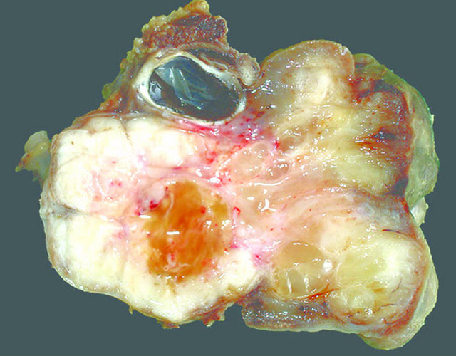
* | * Clusters of edematous and grape-like masses | ||
* Protrusion into [[lumen]] of hollow [[organs]] | |||
[[File:Alveolar rhabdomyosarcoma.jpg|thumb|none|300px| Gross pathology of alveolar rhabdomyosarcoma [http://www.pathologyoutlines.com/wick/softtissue/rhabdomyosarcomaalveolartypeleggross.jpg Source:Courtesy of Mark R. Wick, M.D., from Pathologyoutlines]]] | | style="background:#F5F5F5;" + | | ||
|- | |||
| style="background:#DCDCDC;" align="left" + | | |||
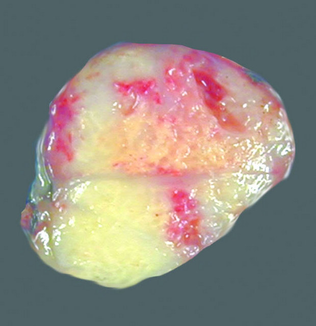
* Poorly circumscribed [[mass]] | |||
* white and infiltrative | |||
* soft or firm [[mass]] | |||
| style="background:#F5F5F5;" + |[[File:Embryonal rhabdomyosarcoma.jpg|thumb|none|300px| Gross feature of orbital embryonal rhabdomyosarcoma [http://www.pathologyoutlines.com/topic/softtissueembryonalrhabdo.html Source: Contributed by Mark R. Wick, M.D., from Pathologyoutlines]]] | |||
|- | |||
| style="background:#DCDCDC;" align="left" + | | |||
*Fleshy [[tumor]] | |||
*Tan gray color [[tumor]] | |||
| style="background:#F5F5F5;" + |[[File:Alveolar rhabdomyosarcoma.jpg|thumb|none|300px| Gross pathology of alveolar rhabdomyosarcoma [http://www.pathologyoutlines.com/wick/softtissue/rhabdomyosarcomaalveolartypeleggross.jpg Source:Courtesy of Mark R. Wick, M.D., from Pathologyoutlines]]] | |||
|} | |||
==Microscopic Pathology== | ==Microscopic Pathology== | ||
=== | {| class="wikitable" | ||
|+ | |||
! style="background:#4479BA; color: #FFFFFF;" align="center" + |Pathologic findings | |||
* | ! style="background:#4479BA; color: #FFFFFF;" align="center" + |Microscopic pathology | ||
* | |- | ||
| style="background:#DCDCDC;" align="left" + | | |||
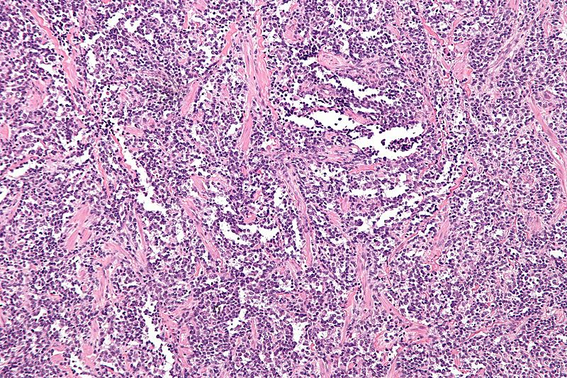
The [[microscopic]] features of [[ARMS]] are:<ref name="pmid15536621">{{cite journal| author=Hostein I, Andraud-Fregeville M, Guillou L, Terrier-Lacombe MJ, Deminière C, Ranchère D et al.| title=Rhabdomyosarcoma: value of myogenin expression analysis and molecular testing in diagnosing the alveolar subtype: an analysis of 109 paraffin-embedded specimens. | journal=Cancer | year= 2004 | volume= 101 | issue= 12 | pages= 2817-24 | pmid=15536621 | doi=10.1002/cncr.20711 | pmc= | url=https://www.ncbi.nlm.nih.gov/entrez/eutils/elink.fcgi?dbfrom=pubmed&tool=sumsearch.org/cite&retmode=ref&cmd=prlinks&id=15536621 }} </ref> | |||
[[Image:800px-Alveolar rhabdomyosarcoma - intermed mag.jpg|thumb|none|300px|Alveolar Rhabdomyosarcoma- intermediate magnification [https://commons.wikimedia.org/wiki/File:Alveolar_rhabdomyosarcoma_-_intermed_mag.jpg Source: Nephron,from Wikimedia Commons]]] | * Ovoid to round [[tumor]] cells are packed with fibrovascular septae | ||
* [[Tumor]] [[cells]] have club-shaped [[appearance]] | |||
* [[Tumor]] [[cells]] are divided by pseudo-alveolar spaces, slightly like [[pulmonary]] [[alveoli]] | |||
* Rhabdomyoblasts are shed into pseudo-alveolar areas | |||
* Presence of more than 50% [[alveolar]] components | |||
| style="background:#F5F5F5;" + |[[Image:800px-Alveolar rhabdomyosarcoma - intermed mag.jpg|thumb|none|300px|Alveolar Rhabdomyosarcoma- intermediate magnification [https://commons.wikimedia.org/wiki/File:Alveolar_rhabdomyosarcoma_-_intermed_mag.jpg Source: Nephron,from Wikimedia Commons]]] | |||
|- | |||
| style="background:#DCDCDC;" align="left" + | | |||
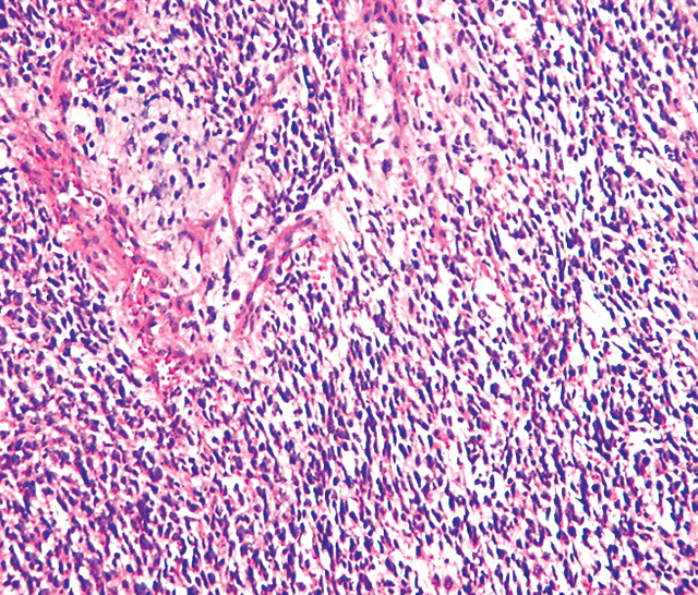
The [[microscopic]] features of ERMS are:<ref name="pmid94079332" /><ref name="pmid9724344">{{cite journal| author=Qualman SJ, Coffin CM, Newton WA, Hojo H, Triche TJ, Parham DM et al.| title=Intergroup Rhabdomyosarcoma Study: update for pathologists. | journal=Pediatr Dev Pathol | year= 1998 | volume= 1 | issue= 6 | pages= 550-61 | pmid=9724344 | doi= | pmc= | url=https://www.ncbi.nlm.nih.gov/entrez/eutils/elink.fcgi?dbfrom=pubmed&tool=sumsearch.org/cite&retmode=ref&cmd=prlinks&id=9724344 }} </ref> | |||
* Rhabdomyoblasts arranged in sheets and large nests | |||
* Different stages of cell [[morphogenesis]] | |||
[[File:Embryonal rhabdomyosarcoma.jpeg|thumb|none|300px| Microscopic pathology of embryonal rhabdomyosarcoma [http://www.pathologyoutlines.com/topic/softtissueembryonalrhabdo.html Source: Contributed by Mark R. Wick, M.D., from Pathologyoutlines]]] | * Positive stains are [[desmin]], [[actin]], [[myogenin]], and MyoD1 | ||
* Infrequent intermixed [[fusiform]] [[cells]] | |||
== | * No [[alveolar]] architectural pattern | ||
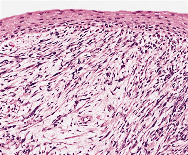
*The microscopic features of botryoid rhabdomyosarcoma:<ref name="pmid26357564">{{cite journal| author=Neha B, Manjunath AP, Girija S, Pratap K| title=Botryoid Rhabdomyosarcoma of the Cervix: Case report with review of the literature. | journal=Sultan Qaboos Univ Med J | year= 2015 | volume= 15 | issue= 3 | pages= e433-7 | pmid=26357564 | doi=10.18295/squmj.2015.15.03.022 | pmc=4554283 | url=https://www.ncbi.nlm.nih.gov/entrez/eutils/elink.fcgi?dbfrom=pubmed&tool=sumsearch.org/cite&retmode=ref&cmd=prlinks&id=26357564 }} </ref> | * Typical rhabdomyoblast has [[eosinophilic]] [[cytoplasm]] with poorly formed myofilaments | ||
** Botryoid rhabdomyosarcoma grows beneath an epithelial surface | | style="background:#F5F5F5;" + |[[File:Embryonal rhabdomyosarcoma.jpeg|thumb|none|300px| Microscopic pathology of embryonal rhabdomyosarcoma [http://www.pathologyoutlines.com/topic/softtissueembryonalrhabdo.html Source: Contributed by Mark R. Wick, M.D., from Pathologyoutlines]]] | ||
** Rhabdomyoblasts adhere to dense subepithelial which is called cambrium layer | |- | ||
**Malignant cells are found in myoxid stroma | | style="background:#DCDCDC;" align="left" + | | ||
[[ File:Botryoid rhabdomyosarcoma.jpg|thumb|none|300px| Microscopic feature of botryoid rhabdomyosarcoma [http://www.pathologyoutlines.com/topic/softtissuebotyroidembryrhabdo.html, from Pathologyoutlines]]] | *The [[microscopic]] features of [[botryoid rhabdomyosarcoma]]:<ref name="pmid26357564">{{cite journal| author=Neha B, Manjunath AP, Girija S, Pratap K| title=Botryoid Rhabdomyosarcoma of the Cervix: Case report with review of the literature. | journal=Sultan Qaboos Univ Med J | year= 2015 | volume= 15 | issue= 3 | pages= e433-7 | pmid=26357564 | doi=10.18295/squmj.2015.15.03.022 | pmc=4554283 | url=https://www.ncbi.nlm.nih.gov/entrez/eutils/elink.fcgi?dbfrom=pubmed&tool=sumsearch.org/cite&retmode=ref&cmd=prlinks&id=26357564 }} </ref> | ||
** [[Botryoid rhabdomyosarcoma]] grows beneath an [[epithelial]] surface | |||
== | ** Rhabdomyoblasts adhere to [[dense]] subepithelial which is called cambrium layer | ||
**[[Malignant]] [[cells]] are found in myoxid [[stroma]] | |||
| style="background:#F5F5F5;" + |[[ File:Botryoid rhabdomyosarcoma.jpg|thumb|none|300px| Microscopic feature of botryoid rhabdomyosarcoma [http://www.pathologyoutlines.com/topic/softtissuebotyroidembryrhabdo.html, from Pathologyoutlines]]] | |||
|- | |||
| style="background:#DCDCDC;" align="left" + | | |||
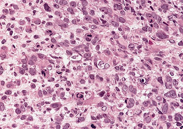
The [[microscopic]] features of [[anaplastic rhabdomyosarcoma]] are:<ref name="pmid24382691">{{cite journal| author=Hettmer S, Archer NM, Somers GR, Novokmet A, Wagers AJ, Diller L et al.| title=Anaplastic rhabdomyosarcoma in TP53 germline mutation carriers. | journal=Cancer | year= 2014 | volume= 120 | issue= 7 | pages= 1068-75 | pmid=24382691 | doi=10.1002/cncr.28507 | pmc=4173134 | url=https://www.ncbi.nlm.nih.gov/entrez/eutils/elink.fcgi?dbfrom=pubmed&tool=sumsearch.org/cite&retmode=ref&cmd=prlinks&id=24382691 }} </ref> | |||
* Presence of large hyperchromatic [[nuclei]] | |||
* Atypical bizarre [[mitotic]] features | |||
* Threefold larger [[nuclear]] size | |||
| style="background:#F5F5F5;" + |[[File:Analastic rhabdomyosarcoma.jpg|thumb|none|300px| Microscopic feature of anaplastic rhabdomyosarcoma [http://www.pathologyoutlines.com/images/softtissue/06_33A.jpg, from Pathologyoutlines]]] | |||
|} | |||
==References== | ==References== | ||
Latest revision as of 16:38, 27 May 2019
|
Rhabdomyosarcoma Microchapters |
|
Diagnosis |
|---|
|
Treatment |
|
Case Studies |
|
Rhabdomyosarcoma pathophysiology On the Web |
|
American Roentgen Ray Society Images of Rhabdomyosarcoma pathophysiology |
|
Risk calculators and risk factors for Rhabdomyosarcoma pathophysiology |
Editor-In-Chief: C. Michael Gibson, M.S., M.D. [1]; Associate Editor(s)-in-Chief: Shadan Mehraban, M.D.[2]
Overview
The origin of rhabdomyosarcoma is straited muscle cells.The presentation sites of rhabdomyosarcoma are head and neck, extremities, Genitourinary tract, trunk, orbit, retroperitoneum, bladder, vagina, nasopharynx, and middle ear.The exact pathogenesis of rhabdomyosarcoma is unclear. However, there is causal association between MET proto-oncogene and macrophage migration inhibitory factor (MIF) and it is thought that P53 is related to oncogenic transformation and tumor progression. Multiple gene mutations are related to rhabdomyosarcoma such as loss of heterozygosity of 11p15, TP53, NRAS, KRAS, HRAS, PIK3CA, CTNNB1, FGFR4, and translocation in PAX3 or PAX7 genes with FOXO1. Immunohistochemistry can identify some specific proteins related to rhabdomyosarcoma such as desmin, actin, myogenin, and myoglobin. The other histologic procedure is transmission electron microscopy (TEM) which can be used for poorly differentiated or undifferentiated tumors and myofilaments, actin, desmin, myotubular intermediate filaments, and rudimentary Z-band material. Fusion protein is identified by reverse transcriptase polymerase chain reaction and fluorescent in situ hybridization.
Pathophysiology
Histology
- The origin of rhabdomyosarcoma is straited muscle cells.[1]
- The presentation sites of rhabdomyosarcoma are:
- Head and neck (28%)
- Extremities (24%)
- Genitourinary tract (18%)
- Trunk (11%)
- Orbit (7%)
- Retroperitoneum (6%)
- Other sites such as bladder, vagina, nasopharynx, and middle ear (3%)
- Round or spindle-shaped muscle cells are present in embryonal rhabdomyosarcoma (ERMS).[2]
- Alveolar architecture with small round undifferentiated cells which are separated by dense hyalinized fibrous septa are present in alveolar rhabdomyosarcoma (ARMS).
- Despite all muscle differentiation markers expression in ARMS, mature muscle characteristics is not seen in ARMS histology.
Pathogenesis
- The exact pathogenesis of rhabdomyosarcoma is unclear.[3]
- Distruption of skeletal muscle progenitor cell growth and related differentiation may cause rhabdomyosarcoma.[3]
- There is causal association between MET proto-oncogene and macrophage migration inhibitory factor (MIF).[4]
- It is thought that P53 is related to oncogenic transformation and tumor progression.[5]
Genetics
- According to microarray and targeted sequencing, following genomic characteristics are attributed to Rhabdomyosarcoma (RMS):[6][7][8][9][10][11]
- Specific genetic changes in different types of rhabdomyosarcoma:
- Alveolar rhabdomyosarcoma:[12][13]
- Translocation of FKHR gene on chromosome 13 with the PAX3 gene on chromosome 2 or the PAX7 gene on chromosome 1
- PAX7 translocation occur in younger patients with longer event-free survival rate
- Embryonal rhabdomyosarcoma:[14]
- Particular gene changes are not clear
- Loss of heterozygosity from the 11p15 region
- Hyperdiploid gene
- Lack of gene amplification
Associated conditions
Immunohistochemistry
- Through immunohistochemistry, muscle specific proteins can be identified in RMS.[2]
- RMS stains positive for following proteins:
Transmission electron microscopy
- Transmission electron microscopy (TEM) can be used for poorly differentiated or undifferentiated tumors.[15][16]
- Presence of following proteins are associated with TEM features of RMS:
Reverse transcriptase polymerase chain reaction (RT-PCR)
Fluorescent in situ hybridization (FISH)
- By using RT-PCR and FISH, features of fusion protein are identified in ARMS.[17]
Gross Pathology
| Gross findings | Gross pathology |
|---|---|
 | |
 |
Microscopic Pathology
| Pathologic findings | Microscopic pathology |
|---|---|
|
The microscopic features of ARMS are:[18] |
 |
|
The microscopic features of ERMS are:[14][19]
|
 |
|
 |
|
The microscopic features of anaplastic rhabdomyosarcoma are:[21] |
 |
References
- ↑ Barr FG (1997). "Molecular genetics and pathogenesis of rhabdomyosarcoma". J Pediatr Hematol Oncol. 19 (6): 483–91. PMID 9407933.
- ↑ 2.0 2.1 Dias P, Chen B, Dilday B, Palmer H, Hosoi H, Singh S; et al. (2000). "Strong immunostaining for myogenin in rhabdomyosarcoma is significantly associated with tumors of the alveolar subclass". Am J Pathol. 156 (2): 399–408. doi:10.1016/S0002-9440(10)64743-8. PMC 1850049. PMID 10666368.
- ↑ 3.0 3.1 Taulli R, Scuoppo C, Bersani F, Accornero P, Forni PE, Miretti S; et al. (2006). "Validation of met as a therapeutic target in alveolar and embryonal rhabdomyosarcoma". Cancer Res. 66 (9): 4742–9. doi:10.1158/0008-5472.CAN-05-4292. PMID 16651427.
- ↑ Tarnowski M, Grymula K, Liu R, Tarnowska J, Drukala J, Ratajczak J; et al. (2010). "Macrophage migration inhibitory factor is secreted by rhabdomyosarcoma cells, modulates tumor metastasis by binding to CXCR4 and CXCR7 receptors and inhibits recruitment of cancer-associated fibroblasts". Mol Cancer Res. 8 (10): 1328–43. doi:10.1158/1541-7786.MCR-10-0288. PMC 2974061. PMID 20861157.
- ↑ Xu J, Timares L, Heilpern C, Weng Z, Li C, Xu H; et al. (2010). "Targeting wild-type and mutant p53 with small molecule CP-31398 blocks the growth of rhabdomyosarcoma by inducing reactive oxygen species-dependent apoptosis". Cancer Res. 70 (16): 6566–76. doi:10.1158/0008-5472.CAN-10-0942. PMC 2922473. PMID 20682800.
- ↑ Scrable H, Cavenee W, Ghavimi F, Lovell M, Morgan K, Sapienza C (1989). "A model for embryonal rhabdomyosarcoma tumorigenesis that involves genome imprinting". Proc Natl Acad Sci U S A. 86 (19): 7480–4. PMC 298088. PMID 2798419.
- ↑ Taylor AC, Shu L, Danks MK, Poquette CA, Shetty S, Thayer MJ; et al. (2000). "P53 mutation and MDM2 amplification frequency in pediatric rhabdomyosarcoma tumors and cell lines". Med Pediatr Oncol. 35 (2): 96–103. PMID 10918230.
- ↑ Stratton MR, Fisher C, Gusterson BA, Cooper CS (1989). "Detection of point mutations in N-ras and K-ras genes of human embryonal rhabdomyosarcomas using oligonucleotide probes and the polymerase chain reaction". Cancer Res. 49 (22): 6324–7. PMID 2680062.
- ↑ Shukla N, Ameur N, Yilmaz I, Nafa K, Lau CY, Marchetti A; et al. (2012). "Oncogene mutation profiling of pediatric solid tumors reveals significant subsets of embryonal rhabdomyosarcoma and neuroblastoma with mutated genes in growth signaling pathways". Clin Cancer Res. 18 (3): 748–57. doi:10.1158/1078-0432.CCR-11-2056. PMC 3271129. PMID 22142829.
- ↑ Taylor JG, Cheuk AT, Tsang PS, Chung JY, Song YK, Desai K; et al. (2009). "Identification of FGFR4-activating mutations in human rhabdomyosarcomas that promote metastasis in xenotransplanted models". J Clin Invest. 119 (11): 3395–407. doi:10.1172/JCI39703. PMC 2769177. PMID 19809159.
- ↑ Barr FG, Galili N, Holick J, Biegel JA, Rovera G, Emanuel BS (1993). "Rearrangement of the PAX3 paired box gene in the paediatric solid tumour alveolar rhabdomyosarcoma". Nat Genet. 3 (2): 113–7. doi:10.1038/ng0293-113. PMID 8098985.
- ↑ Missiaglia E, Williamson D, Chisholm J, Wirapati P, Pierron G, Petel F; et al. (2012). "PAX3/FOXO1 fusion gene status is the key prognostic molecular marker in rhabdomyosarcoma and significantly improves current risk stratification". J Clin Oncol. 30 (14): 1670–7. doi:10.1200/JCO.2011.38.5591. PMID 22454413.
- ↑ Cao L, Yu Y, Bilke S, Walker RL, Mayeenuddin LH, Azorsa DO; et al. (2010). "Genome-wide identification of PAX3-FKHR binding sites in rhabdomyosarcoma reveals candidate target genes important for development and cancer". Cancer Res. 70 (16): 6497–508. doi:10.1158/0008-5472.CAN-10-0582. PMC 2922412. PMID 20663909.
- ↑ 14.0 14.1
- ↑ Hicks J, Flaitz C (2002). "Rhabdomyosarcoma of the head and neck in children". Oral Oncol. 38 (5): 450–9. PMID 12110339.
- ↑ Parham DM (2001). "Pathologic classification of rhabdomyosarcomas and correlations with molecular studies". Mod Pathol. 14 (5): 506–14. doi:10.1038/modpathol.3880339. PMID 11353062.
- ↑ Helman LJ, Meltzer P (2003). "Mechanisms of sarcoma development". Nat Rev Cancer. 3 (9): 685–94. doi:10.1038/nrc1168. PMID 12951587.
- ↑ Hostein I, Andraud-Fregeville M, Guillou L, Terrier-Lacombe MJ, Deminière C, Ranchère D; et al. (2004). "Rhabdomyosarcoma: value of myogenin expression analysis and molecular testing in diagnosing the alveolar subtype: an analysis of 109 paraffin-embedded specimens". Cancer. 101 (12): 2817–24. doi:10.1002/cncr.20711. PMID 15536621.
- ↑ Qualman SJ, Coffin CM, Newton WA, Hojo H, Triche TJ, Parham DM; et al. (1998). "Intergroup Rhabdomyosarcoma Study: update for pathologists". Pediatr Dev Pathol. 1 (6): 550–61. PMID 9724344.
- ↑ Neha B, Manjunath AP, Girija S, Pratap K (2015). "Botryoid Rhabdomyosarcoma of the Cervix: Case report with review of the literature". Sultan Qaboos Univ Med J. 15 (3): e433–7. doi:10.18295/squmj.2015.15.03.022. PMC 4554283. PMID 26357564.
- ↑ Hettmer S, Archer NM, Somers GR, Novokmet A, Wagers AJ, Diller L; et al. (2014). "Anaplastic rhabdomyosarcoma in TP53 germline mutation carriers". Cancer. 120 (7): 1068–75. doi:10.1002/cncr.28507. PMC 4173134. PMID 24382691.