Blepharitis other diagnostic studies: Difference between revisions
m (→References) |
No edit summary |
||
| Line 38: | Line 38: | ||
===Biopsy=== | ===Biopsy=== | ||
Biopsy of the eyelid may be indicated in the following conditions: | Biopsy of the eyelid may be indicated in the following conditions: | ||
*Blepharitis with marked | *Blepharitis with marked asymmetry (possibility of [[carcinoma]]) | ||
*Blepharitis not responding to therapy | *Blepharitis not responding to therapy | ||
*Unifocal recurrent blepharitis with [[chalazion|chalazia]] | *Unifocal recurrent blepharitis with [[chalazion|chalazia]] | ||
Revision as of 19:37, 27 July 2016
|
Blepharitis Microchapters | |
|
Diagnosis | |
|---|---|
|
Treatment | |
|
Case Studies | |
|
Blepharitis other diagnostic studies On the Web | |
|
American Roentgen Ray Society Images of Blepharitis other diagnostic studies | |
|
Risk calculators and risk factors for Blepharitis other diagnostic studies | |
Editor-In-Chief: C. Michael Gibson, M.S., M.D. [1]; Associate Editor(s)-in-Chief: Sara Mehrsefat, M.D. [2]
Overview
Other diagnostic studies for blepharitis include slit lamp examination, tear break up time (TBUT), and measurement of tear osmolarity.[1][2][3]
Other Diagnostic Studies
Other diagnostic studies for blepharitis include:[1][2][3][4]
Slit lamp examination
On slit lamp examination, blepharitis is characterized by:
- Meibomian gland enlargement
- Waxy secretions at the gland openings
- Lid margin neovascularization and dilation of existing blood vessels
- Ulcerations along the eyelid margin
- Corneal nodules
- Corneal margin ulcer
- Superficial corneal pannus
Images
The following are slit lamp examination images associated with blepharitis:[1]
-
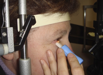
Gland expression performed at the slit lamp
-
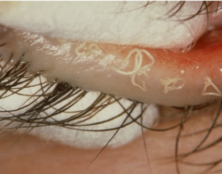
Waxy secretions at the gland openings
-
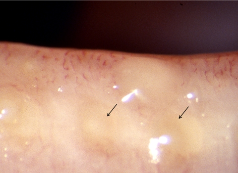
Cloudy expressed meibum (arrows)


Tear Break Up Time
On tear break up time (TBUT), blepharitis is characterized by tear film instability and rapid evaporation.
Measurement of Tear Osmolarity
On measurement of tear osmolarity, blepharitis concurrent with dry eye syndrome may be characterized by tear osmolarity of 316 mOsm/L or greater.
Microscopic evaluation of eyelashes
On microscopic evaluation of eyelashes, chronic blepharoconjunctivitis may be characterized by the presence of Demodex mites.
Biopsy
Biopsy of the eyelid may be indicated in the following conditions:
- Blepharitis with marked asymmetry (possibility of carcinoma)
- Blepharitis not responding to therapy
- Unifocal recurrent blepharitis with chalazia
References
- ↑ 1.0 1.1 1.2 Tomlinson A, Bron AJ, Korb DR, Amano S, Paugh JR, Pearce EI; et al. (2011). "The international workshop on meibomian gland dysfunction: report of the diagnosis subcommittee". Invest Ophthalmol Vis Sci. 52 (4): 2006–49. doi:10.1167/iovs.10-6997f. PMC 3072162. PMID 21450918.
- ↑ 2.0 2.1 Driver PJ, Lemp MA (1996). "Meibomian gland dysfunction". Surv Ophthalmol. 40 (5): 343–67. PMID 8779082.
- ↑ 3.0 3.1 Bachmeyer C, Bégon E (2013). "Chronic blepharitis". Neth J Med. 71 (5): 259–63. PMID 23799315.
- ↑ McCulley JP, Dougherty JM (1985). "Blepharitis associated with acne rosacea and seborrheic dermatitis". Int Ophthalmol Clin. 25 (1): 159–72. PMID 3156100.