Astrocytoma pathophysiology
|
Astrocytoma Microchapters |
|
Diagnosis |
|---|
|
Treatment |
|
Case Study |
|
Astrocytoma pathophysiology On the Web |
|
American Roentgen Ray Society Images of Astrocytoma pathophysiology |
|
Risk calculators and risk factors for Astrocytoma pathophysiology |
Editor-In-Chief: C. Michael Gibson, M.S., M.D. [1];Associate Editor(s)-in-Chief: Shivali Marketkar, M.B.B.S. [2]; Ammu Susheela, M.D. [3]
Overview
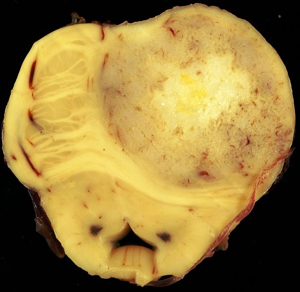
On gross pathology, compression, invasion and destruction of brain parenchyma are characteristic findings of astrocytoma. On microscopic histopathological analysis, gemistocytes, rosenthal fibres and hyalinisation of blood vessels are characteristic findings of astrocytoma.
Pathophysiology
Gross Pathology

- Astrocytoma causes regional effects by compression, invasion, and destruction of brain parenchyma, arterial and venous hypoxia, competition for nutrients, release of metabolic end products (e.g., free radicals, altered electrolytes, neurotransmitters), and release and recruitment of cellular mediators e.g., cytokines) that disrupt normal parenchymal function. Secondary clinical sequelae may be caused by elevated intracranial pressure (ICP) attributable to direct mass effect, increased blood volume, or increased cerebrospinal fluid (CSF) volume.[1]
Microscopic Pathology
- Histologic diagnosis with tissue biopsy will normally reveal an infiltrative character suggestive of the slow growing nature of the tumor. The tumor may be cavitating, pseudocyst-forming, or noncavitating. Appearance is usually white-gray, firm, and almost indistinguishable from normal white matter.
Histopathological Video
Video
{{#ev:youtube|O0b4zyDQcyI}}