Aspergillosis chest x ray: Difference between revisions
No edit summary |
m (Changes made per Mahshid's request) |
||
| (18 intermediate revisions by 4 users not shown) | |||
| Line 1: | Line 1: | ||
__NOTOC__ | |||
{{Aspergillosis}} | {{Aspergillosis}} | ||
{{CMG}}; | {{CMG}} {{AE}} {{HL}}; {{YD}}; {{SSK}} | ||
==Overview== | ==Overview== | ||
Chest X-ray may be helpful in the diagnosis of aspergillosis. Chest X-ray may be remarkable for recurrent pulmonary infiltrates in allergic bronchopulmonary aspergillosis. Findings on chest X-ray suggestive of aspergilloma include a well demarcated, round, soft tissue mass located inside an air space cavity of the [[lung]]s.<ref>{{cite journal | author = Curtis A, Smith G, Ravin C | title = Air crescent sign of invasive aspergillosis. | journal = Radiology | volume = 133 | issue = 1 | pages = 17-21 | year = 1979 | month = Oct | id = PMID 472287 | url=http://radiology.rsnajnls.org/cgi/content/abstract/133/1/17?ijkey=657da61e6546d66bd27550542bc4d25be2efe638&keytype2=tf_ipsecsha}}</ref><ref name="radio">Aspergilloma. Radiopaedia (2015) http://radiopaedia.org/articles/aspergilloma Accessed on February, 9 2016</ref> Multiple cavities with evidence of fibrosis are suggestive of chronic pulmonary aspergillosis, whereas focal nodules and infiltration are suggestive of invasive aspergillosis. | |||
==Chest X-Ray== | |||
* Chest X-ray may be helpful in the diagnosis of aspergillosis. | |||
* Findings on [[chest X-ray]] suggestive of aspergillosis include: | |||
:* A well demarcated, round, soft tissue mass | |||
:* Located inside an air space cavity of the [[lung]]s | |||
:* [[Air crescent sign]] | |||
:* Altering the position of the patient usually demonstrates that the mass is mobile | |||
==Chest X-ray | ==Gallery== | ||
<gallery> | |||
Image: | |||
Aspergillosis X-ray.jpg|Chest X-ray of a patient demonstrates a rounded opacity located at the medial aspect of the right lung apex | |||
Image: | |||
Aspergillosis X-ray 2.jpg|A close up view on the patient demonstrates a well circumscribed, rounded opacity located at the medial aspect of the right lung apex | |||
Image: | |||
Aspergillosis X-ray 3.jpg|Chest X-ray of a patient demonstrates a rounded soft tissue attenuating masses located inside a cavitary lesion observed at the middle lobe of the right lung | |||
Image: | |||
Aspergillosis X-ray 4.jpg|Air cresent sign aspergillosis | |||
Image: | |||
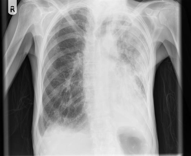
Aspergillosis X-ray 7.jpg|Chest X-ray that demonstrate a densitiy in the right upper zone with clear evidence of volume loss (the trachea and mediastinum are pulled towards the right, and the hilum is elevated) | |||
</gallery> | |||
==References== | ==References== | ||
| Line 12: | Line 31: | ||
[[Category:Fungal diseases]] | [[Category:Fungal diseases]] | ||
} | } | ||
{{WH}} | {{WH}} | ||
{{WS}} | {{WS}} | ||
Latest revision as of 16:44, 18 September 2017
|
Aspergillosis Microchapters |
|
Diagnosis |
|---|
|
Treatment |
|
Case Studies |
|
Aspergillosis chest x ray On the Web |
|
American Roentgen Ray Society Images of Aspergillosis chest x ray |
|
Risk calculators and risk factors for Aspergillosis chest x ray |
Editor-In-Chief: C. Michael Gibson, M.S., M.D. [1] Associate Editor(s)-in-Chief: Haytham Allaham, M.D. [2]; Yazan Daaboul, M.D.; Serge Korjian M.D.
Overview
Chest X-ray may be helpful in the diagnosis of aspergillosis. Chest X-ray may be remarkable for recurrent pulmonary infiltrates in allergic bronchopulmonary aspergillosis. Findings on chest X-ray suggestive of aspergilloma include a well demarcated, round, soft tissue mass located inside an air space cavity of the lungs.[1][2] Multiple cavities with evidence of fibrosis are suggestive of chronic pulmonary aspergillosis, whereas focal nodules and infiltration are suggestive of invasive aspergillosis.
Chest X-Ray
- Chest X-ray may be helpful in the diagnosis of aspergillosis.
- Findings on chest X-ray suggestive of aspergillosis include:
- A well demarcated, round, soft tissue mass
- Located inside an air space cavity of the lungs
- Air crescent sign
- Altering the position of the patient usually demonstrates that the mass is mobile
Gallery
-
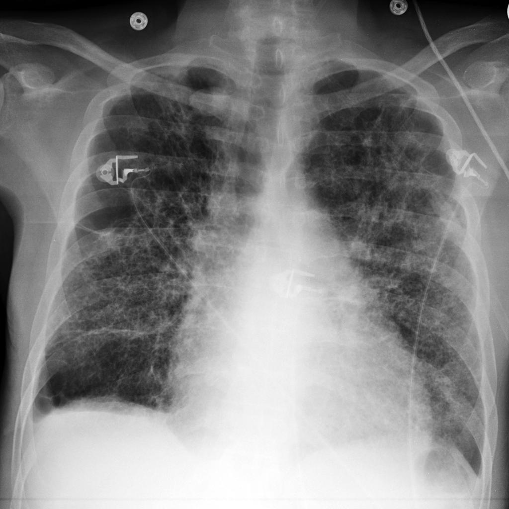
Chest X-ray of a patient demonstrates a rounded opacity located at the medial aspect of the right lung apex
-
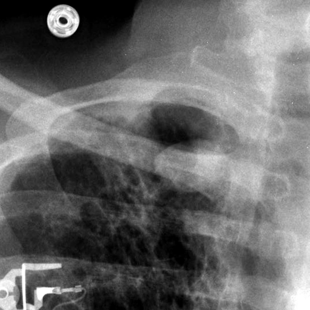
A close up view on the patient demonstrates a well circumscribed, rounded opacity located at the medial aspect of the right lung apex
-
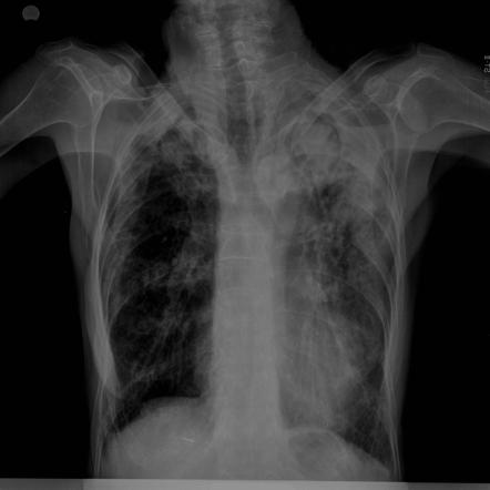
Chest X-ray of a patient demonstrates a rounded soft tissue attenuating masses located inside a cavitary lesion observed at the middle lobe of the right lung
-
Air cresent sign aspergillosis
-
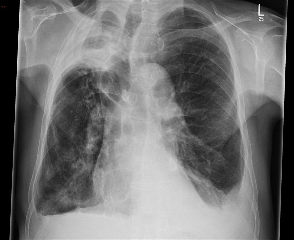
Chest X-ray that demonstrate a densitiy in the right upper zone with clear evidence of volume loss (the trachea and mediastinum are pulled towards the right, and the hilum is elevated)


References
- ↑ Curtis A, Smith G, Ravin C (1979). "Air crescent sign of invasive aspergillosis". Radiology. 133 (1): 17–21. PMID 472287. Unknown parameter
|month=ignored (help) - ↑ Aspergilloma. Radiopaedia (2015) http://radiopaedia.org/articles/aspergilloma Accessed on February, 9 2016