Acute bronchitis pathophysiology: Difference between revisions
Jump to navigation
Jump to search
m (Bot: Removing from Primary care) |
|||
| (8 intermediate revisions by 4 users not shown) | |||
| Line 3: | Line 3: | ||
==Overview== | ==Overview== | ||
The pathologic process starts with the inoculation of tracheobranchial [[epithelium]] with invading organism, which leads to [[inflammation]], thickening and increased [[mucus]] production. | |||
==Pathophysiology== | ==Pathophysiology== | ||
*The causative agent | ===Pathogenesis=== | ||
*Following transmission, the | *The causative agent is transmitted through the large and medium size airway tracts.<ref name="pmid11119400">{{cite journal |vauthors=Gonzales R, Sande MA |title=Uncomplicated acute bronchitis |journal=Ann. Intern. Med. |volume=133 |issue=12 |pages=981–91 |year=2000 |pmid=11119400 |doi= |url=}}</ref> | ||
*This process leads to inflammation, thickening, and increased mucus production in the airways as shown | *Following transmission, the agent inoculates the tracheobronchial [[epithelium]]. | ||
*This process leads to [[inflammation]], thickening, and increased [[mucus]] production in the airways compared to normal [[bronchi]] as shown below: | |||
<gallery> | <gallery> | ||
Image:normal bronchi.jpg| | Image:normal bronchi.jpg|Normal Bronchi | ||
Image:acute bronchitis.jpg| | Image:acute bronchitis.jpg|Inflamed Bronchi | ||
</gallery> | </gallery> | ||
===Microscopy=== | |||
*On microscopic analysis, [[Epithelial cell|epithelial-cell]] desquamation and denuding of the airway to the level of the [[basement membrane]], in association with the presence of a [[lymphocytic]] cellular infiltrate, have been demonstrated.<ref name="pmid13782910">{{cite journal |vauthors=WALSH JJ, DIETLEIN LF, LOW FN, BURCH GE, MOGABGAB WJ |title=Bronchotracheal response in human influenza. Type A, Asian strain, as studied by light and electron microscopic examination of bronchoscopic biopsies |journal=Arch. Intern. Med. |volume=108 |issue= |pages=376–88 |year=1961 |pmid=13782910 |doi= |url=}}</ref> | |||
==References== | ==References== | ||
| Line 25: | Line 28: | ||
{{WikiDoc Sources}} | {{WikiDoc Sources}} | ||
[[Category:Pulmonology]] | [[Category:Pulmonology]] | ||
[[Category:Disease]] | [[Category:Disease]] | ||
[[Category:Surgery]] | |||
[[Category:Up-To-Date]] | |||
[[Category:Emergency medicine]] | |||
[[Category:Infectious disease]] | |||
Latest revision as of 20:15, 29 July 2020
|
Acute bronchitis Microchapters |
|
Diagnosis |
|
Treatment |
|
Acute bronchitis pathophysiology On the Web |
|
American Roentgen Ray Society Images of Acute bronchitis pathophysiology |
|
Risk calculators and risk factors for Acute bronchitis pathophysiology |
Editor-In-Chief: C. Michael Gibson, M.S., M.D. [1]Associate Editor(s)-in-Chief: Seyedmahdi Pahlavani, M.D. [2]
Overview
The pathologic process starts with the inoculation of tracheobranchial epithelium with invading organism, which leads to inflammation, thickening and increased mucus production.
Pathophysiology
Pathogenesis
- The causative agent is transmitted through the large and medium size airway tracts.[1]
- Following transmission, the agent inoculates the tracheobronchial epithelium.
- This process leads to inflammation, thickening, and increased mucus production in the airways compared to normal bronchi as shown below:
-
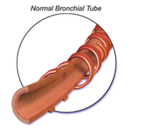
Normal Bronchi
-
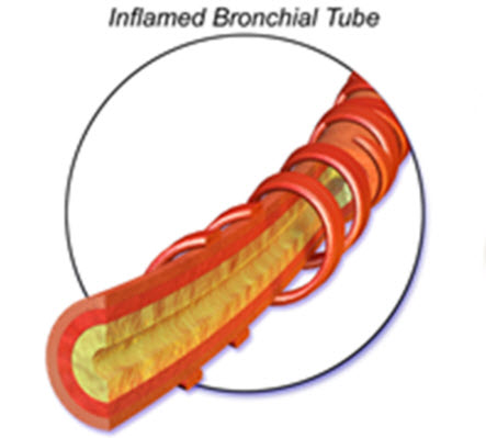
Inflamed Bronchi


Microscopy
- On microscopic analysis, epithelial-cell desquamation and denuding of the airway to the level of the basement membrane, in association with the presence of a lymphocytic cellular infiltrate, have been demonstrated.[2]
References
- ↑ Gonzales R, Sande MA (2000). "Uncomplicated acute bronchitis". Ann. Intern. Med. 133 (12): 981–91. PMID 11119400.
- ↑ WALSH JJ, DIETLEIN LF, LOW FN, BURCH GE, MOGABGAB WJ (1961). "Bronchotracheal response in human influenza. Type A, Asian strain, as studied by light and electron microscopic examination of bronchoscopic biopsies". Arch. Intern. Med. 108: 376–88. PMID 13782910.