Wolff-Parkinson-White syndrome EKG examples
Editor-In-Chief: C. Michael Gibson, M.S., M.D. [1]
For the main page on Wolff-Parkinson-White syndrome click here
EKG examples
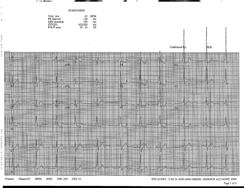
Shown below is an electrocardiogram of Wolff-Parkinson-White syndrome.

Shown below is an electrocardiogram of Wolff-Parkinson-White syndrome.

Shown below is an electrocardiogram of Wolff-Parkinson-White syndrome (antero-lateral pathway).

Shown below is an electrocardiogram of Wolff-Parkinson-White syndrome depicting delta wave.

Shown below is an electrocardiogram of Wolff-Parkinson-White syndrome (antero-septal pathway).

Shown below is an electrocardiogram of Wolff-Parkinson-White syndrome (antero-septal pathway).

Shown below is an electrocardiogram of Wolff-Parkinson-White syndrome (epicardial pathway).

-
Wolf Parkinson White Left Posterior Pathway
-
Wolf Parkinson White Syndrome Posteroseptal Pathway

Shown below is an EKG showing abnormal QRS form with delta waves seen best in the V leads.

-
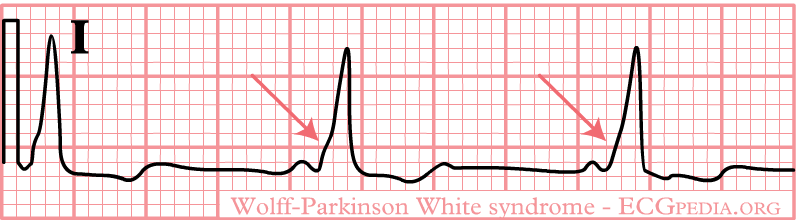
The upstroke of the QRS-complex is 'slurred', resulting in a delta-wave (arrow).
-
Delta waves in a patient with Wolff-Parkinson-White Syndrome (WPW)
-
WPW on a 12 lead ECG.
-
Another example of WPW on a 12 lead ECG.
-
12 lead EKG: Wolff Parkinson White Syndrome
-
12 lead EKG: Wolff Parkinson White Syndrome Type I. Courtesy of Dr Jose Ganseman
-
12 lead EKG: Wolff Parkinson White Syndrome Type I. Courtesy of Dr Jose Ganseman
-
12 lead EKG: Wolff Parkinson White Syndrome Type I. Courtesy of Dr Jose Ganseman
-
12 lead EKG: Wolff Parkinson White Syndrome Type II. Courtesy of Dr Jose Ganseman
-
12 lead EKG: Wolff Parkinson White Syndrome Type II. Courtesy of Dr Jose Ganseman
-
12 lead EKG: Wolff Parkinson White Syndrome Type II. Courtesy of Dr Jose Ganseman
-
12 lead EKG: Wolff Parkinson White Syndrome Type II. Courtesy of Dr Jose Ganseman
-
12 lead EKG: Wolff Parkinson White Syndrome Type II. Courtesy of Dr Jose Ganseman
-
WPW syndrome with an orthodromic circus movement tachycardia: Narrow complex tachycardia with a rate of 200 bpm (RR interval 320 ms). After 5 cycles, the tachycardia suddenly stops and four multiform complexes are seen without any P waves. These complexes should be regarded as a polymorphic ventricular tachycardia, which is not uncommon after an adenosine-terminated supraventricular tachycardia. A 5th complex is preceded by a P wave. The subsequent 4 complexes show a widened QRS complex and all are immediately preceded by a P wave. The initial phase of the QRS complex is slurred and positive in all available leads. Sinus rhythm continues thereafter with gradual abbreviation of the QRS complex until a 120 msec wide QRS complex remains.
-
The same patient's EKG during sinus rhythm. A discrete Δ wave is clearly visible. The morphology of the Δ wave suggests a left posterior Kent bundle.
Sources
Copyleft images obtained courtesy of ECGpedia, http://en.ecgpedia.org/index.php?title=Special:NewFiles&offset=&limit=500