Yersinia pestis infection causes
|
Yersinia pestis infection Microchapters |
|
Differentiating Yersinia Pestis Infection from other Diseases |
|---|
|
Diagnosis |
|
Treatment |
|
Case Studies |
|
Yersinia pestis infection causes On the Web |
|
American Roentgen Ray Society Images of Yersinia pestis infection causes |
|
Risk calculators and risk factors for Yersinia pestis infection causes |
Editor-In-Chief: C. Michael Gibson, M.S., M.D. [1]; Assistant Editors-In-Chief: Esther Lee, M.A.; Rim Halaby, M.D. [2]
Overview
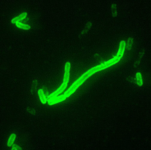
Yersinia pestis (Y. pestis), a rod-shaped facultative anaerobe with bipolar staining (giving it a safety pin appearance) causes the infection in mammals and humans.[1] The bacteria maintain their existence in a cycle involving rodents and their fleas. The genus Yersinia is gram-negative, bipolar staining coccobacilli, and, similarly to other Enterobacteriaceae, it has a fermentative metabolism. Y. pestis produces an antiphagocytic slime. The organism is motile when isolated, but becomes nonmotile in the mammalian host.
Taxonomy
Bacteria; Proteobacteria; Gammaproteobacteria; Enterobacteriales; Yersinia; Yersinia pestis
Biology
Yersinia pests is a nonmotile, non-spore-forming, Gram-negative, non-lactose fermenting, bipolar, ovoid, "safety-pin-shaped" bacillus.
Genome
The complete genomic sequence is available for two of the three sub-species of yersinia pestis: strain KIM (of biovar Medievalis),[2] and strain CO92 (of biovar Orientalis, obtained from a clinical isolate in the United States).[3] As of 2006, the genomic sequence of a strain of biovar Antiqua has been recently completed.[4] Similar to the other pathogenic strains, there are signs of loss of function mutations. The chromosome of strain KIM is 4,600,755 base pairs long; the chromosome of strain CO92 is 4,653,728 base pairs long. Like its cousins Yersinia pseudotuberculosis and Yersinia enterocolitica, Yersinia pestis is host to the plasmid pCD1. In addition, it also hosts two other plasmids, pPCP1 (also called pPla or pPst) and pMT1 (also called pFra) that are not carried by the other Yersinia species. pFra codes for a phospholipase D that is important for the ability of Yersinia pestis to be transmitted by fleas. pPla codes for a protease, Pla, that activates plasminogen in human hosts and is a very important virulence factor for pneumonic plague.[5] Together, these plasmids, and a pathogenicity island called HPI, encode several proteins that cause the pathogenesis, for which Yersinia pestis is famous. Among other things, these virulence factors are required for bacterial adhesion and injection of proteins into the host cell, invasion of bacteria in the host cell (via a Type III secretion system), and acquisition and binding of iron that is harvested from red blood cells (via siderophores). Yersinia pestis is thought to be descendant from Yersinia pseudotuberculosis, differing only in the presence of specific virulence plasmids.
A comprehensive and comparative proteomics analysis of Yersinia pestis strain KIM was performed in 2006.[6] The analysis focused on the transition to a growth condition mimicking growth in host cells.
Tropism
Natural reservoir
References
- ↑ Collins FM (1996). Pasteurella, Yersinia, and Francisella. In: Baron's Medical Microbiology (Baron S et al, eds.) (4th ed.). Univ. of Texas Medical Branch. ISBN 0-9631172-1-1.
- ↑ Deng W; et al. (2002). "Genome Sequence of Yersinia pestis KIM". Journal of Bacteriology. 184 (16): 4601&ndash, 4611. doi:10.1128/JB.184.16.4601-4611.2002. PMC 135232. PMID 12142430. Unknown parameter
|author-separator=ignored (help) - ↑ Parkhill J; et al. (2001). "Genome sequence of Yersinia pestis, the causative agent of plague". Nature. 413 (6855): 523&ndash, 527. doi:10.1038/35097083. PMID 11586360. Unknown parameter
|author-separator=ignored (help) - ↑ Chain PS; Hu P; Malfatti SA; et al. (2006). "Complete Genome Sequence of Yersinia pestis Strains Antiqua and Nepal516: Evidence of Gene Reduction in an Emerging Pathogen". J. Bacteriol. 188 (12): 4453–63. doi:10.1128/JB.00124-06. PMC 1482938. PMID 16740952. Unknown parameter
|author-separator=ignored (help) - ↑ Lathem WW, Price PA, Miller VL, Goldman WE (2007). "A plasminogen-activating protease specifically controls the development of primary pneumonic plague". Science. 315 (5811): 509–13. doi:10.1126/science.1137195. PMID 17255510.
- ↑ Hixson K; et al. (2006). "Biomarker candidate identification in Yersinia pestis using organism-wide semiquantitative proteomics". Journal of Proteome Research. 5 (11): 3008–3017. doi:10.1021/pr060179y. PMID 16684765. Unknown parameter
|author-separator=ignored (help)

