Burkitt's lymphoma pathophysiology: Difference between revisions
No edit summary |
No edit summary |
||
| Line 1: | Line 1: | ||
__NOTOC__ | __NOTOC__ | ||
{{Burkitt's lymphoma}} | {{Burkitt's lymphoma}} | ||
{{CMG}};{{SM}} | |||
{{CMG}} | |||
==Overview== | ==Overview== | ||
| Line 23: | Line 21: | ||
[[Image:800px-Burkitt_lymphoma,_touch_prep,_Wright_stain.jpg|200px]] | [[Image:800px-Burkitt_lymphoma,_touch_prep,_Wright_stain.jpg|200px]] | ||
| Line 62: | Line 59: | ||
[[Category:Types of cancer]] | [[Category:Types of cancer]] | ||
[[Category:Oncology]] | [[Category:Oncology]] | ||
[[Category:Needs | [[Category:Needs overview]] | ||
Revision as of 12:08, 11 March 2013
|
Burkitt's lymphoma Microchapters |
|
Diagnosis |
|---|
|
Treatment |
|
Case Studies |
|
Burkitt's lymphoma pathophysiology On the Web |
|
American Roentgen Ray Society Images of Burkitt's lymphoma pathophysiology |
|
Risk calculators and risk factors for Burkitt's lymphoma pathophysiology |
Editor-In-Chief: C. Michael Gibson, M.S., M.D. [1];Shivali Marketkar, M.B.B.S. [2]
Overview
Pathophysiology
Gross Pathology
-
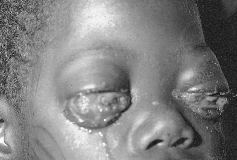
Burkitt lymphoma: bilateral eye involvement

Microscopic Pathology
The tumor consists of sheets of a monotonous (i.e. similar in size and morphology) population of medium size lymphoid cells with high proliferative activity and apoptotic activity. The "starry sky" appearance seen[1] under low power is due to scattered tingible body-laden macrophages (macrophages containing dead body of apoptotic tumor cells). The old descriptive term of "small non-cleaved cell" is misleading. The tumor cells are mostly medium in size (i.e. tumor nuclei size similar to that of histiocytes or endothelial cells). "Small non-cleaved cells" are compared to "large non-cleaved cells" of normal germinal center lymphocytes. Tumor cells possess small amount of basophilic cytoplasm. The cellular outline usually appears squared off.Shown below is a micrograph of touch prep stained with Wright's stain.

Immunohistochemistry
The tumor cells in Burkitt lymphoma generally strongly express markers of B cell differentiation (CD20, CD22, CD19) as well as CD10, and BCL6. The tumour cells are generally negative for BCL2 and TdT. The high mitotic activity of Burkitt lymphoma is confirmed by nearly 100% of the cells staining positive for Ki67.[2]
Genetics
All types of Burkitt's lymphoma are characterized by disregulation of the c-myc gene by one of three chromosomal translocations.[3] This gene is found at 8q24.
- The most common variant is t(8;14)(q24;q32), which accounts for approximately 85%[3] of cases. This involves c-myc and IGH@. A variant of this, a three-way translocation, t(8;14;18), has also been identified.[4]
Combined, the two less-common translocations, t(2;8)(p12;q24) and t(8;22)(q24;q11), account for the remaining 15% of cases not due to the t(8;14)(q24;q32) translocation.[3]
Malignant B cell characteristics
Malignant B cells have identical DNA recombinations of the V(D)J region of the Immunoglobin genes. This means that no increase in specificity of Antibody molecules is occurring in the malignant cells. These malignant cells are thus clonal populations and can be assayed for by using DNA probes specific for the regions where recombination is expected. Normal DNA will be characterized by two high concentration of identical germ line DNA V(D)J regions and endless, likely undetectable, non-germline Ig V(D)J DNA. Lymphoma cells have an additional high concentration of V(D)J DNA that is unlike the germline, indicating clonal populations of B Cells that are not undifferentiated B Cells (Germline DNA cells). Assays typically use the process of Electrophoresis and southern blot analysis to determine the existence of these characteristics.
References
- ↑ Fujita S, Buziba N, Kumatori A, Senba M, Yamaguchi A, Toriyama K (2004). "Early stage of Epstein-Barr virus lytic infection leading to the "starry sky" pattern formation in endemic Burkitt lymphoma". Arch. Pathol. Lab. Med. 128 (5): 549–52. doi:10.1043/1543-2165(2004)128<549:ESOEVL>2.0.CO;2. PMID 15086279. Unknown parameter
|month=ignored (help) - ↑ . ISBN 978-92-832-2431-0. Missing or empty
|title=(help) - ↑ 3.0 3.1 3.2 Hoffman, Ronald (2009). Hematology : basic principles and practice (PDF) (5th ed. ed.). Philadelphia, PA: Churchill Livingstone/Elsevier. pp. 1304–1305. ISBN 978-0-443-06715-0.
- ↑ Liu D, Shimonov J, Primanneni S, Lai Y, Ahmed T, Seiter K (2007). "t(8;14;18): a 3-way chromosome translocation in two patients with Burkitt's lymphoma/leukemia". Mol. Cancer. 6: 35. doi:10.1186/1476-4598-6-35. PMC 1904237. PMID 17547754.
- ↑ 5.0 5.1 Smardova J, Grochova D, Fabian P; et al. (2008). "An unusual p53 mutation detected in Burkitt's lymphoma: 30 bp duplication". Oncol. Rep. 20 (4): 773–8. PMID 18813817. Unknown parameter
|month=ignored (help)