Wolff-Parkinson-White syndrome EKG examples: Difference between revisions
No edit summary |
|||
| Line 27: | Line 27: | ||
---- | ---- | ||
<div align="left"> | |||
<gallery heights="175" widths="175"> | |||
Image:WPWLP.jpg|Wolf Parkinson White Left Posterior Pathway | |||
Image:WPW.jpg|Wolf Parkinson White Syndrome Posteroseptal Pathway | |||
</gallery> | |||
</div> | |||
---- | |||
Shown below is an EKG showing abnormal QRS form with delta waves seen best in the V leads. | |||
[[File:WPWEKG.jpg|center|800px]] | |||
---- | |||
<div align="left"> | |||
<gallery heights="175" widths="175"> | |||
Image:Rhythm_WPW.png|The upstroke of the QRS-complex is 'slurred', resulting in a delta-wave (arrow). | |||
</gallery> | |||
</div> | |||
<div align="left"> | |||
<gallery heights="175" widths="175"> | |||
image:Wolff-Parkinson-White-syndrome.jpg|Delta waves in a patient with Wolff-Parkinson-White Syndrome (WPW) | |||
</gallery> | |||
</div> | |||
<div align="left"> | |||
<gallery heights="175" widths="175"> | |||
Image:wpw_full_ecg.jpg|WPW on a 12 lead ECG. | |||
Image:wpw_full_ecg2.jpg|Another example of WPW on a 12 lead ECG. | |||
</gallery> | |||
</div> | |||
<div align="left"> | |||
<gallery heights="175" widths="175"> | |||
Image:wpw_full_ecg3.png|12 lead EKG: Wolff Parkinson White Syndrome | |||
Image:Ganseman WPW type 1.1.jpg|12 lead EKG: Wolff Parkinson White Syndrome Type I. [http://www.ganseman.com/ecgbibnl.htm#_top000 Courtesy of Dr Jose Ganseman] | |||
</gallery> | |||
</div> | |||
<div align="left"> | |||
<gallery heights="175" widths="175"> | |||
Image:Ganseman WPW type 1.2.jpg|12 lead EKG: Wolff Parkinson White Syndrome Type I. [http://www.ganseman.com/ecgbibnl.htm#_top000 Courtesy of Dr Jose Ganseman] | |||
Image:Ganseman WPW type 1.3.jpg|12 lead EKG: Wolff Parkinson White Syndrome Type I. [http://www.ganseman.com/ecgbibnl.htm#_top000 Courtesy of Dr Jose Ganseman] | |||
</gallery> | |||
</div> | |||
<div align="left"> | |||
<gallery heights="175" widths="175"> | |||
Image:Ganseman WPW type 2.1.jpg|12 lead EKG: Wolff Parkinson White Syndrome Type II. [http://www.ganseman.com/ecgbibnl.htm#_top000 Courtesy of Dr Jose Ganseman] | |||
Image:Ganseman WPW type 2.2.jpg|12 lead EKG: Wolff Parkinson White Syndrome Type II. [http://www.ganseman.com/ecgbibnl.htm#_top000 Courtesy of Dr Jose Ganseman] | |||
</gallery> | |||
</div> | |||
<div align="left"> | |||
<gallery heights="175" widths="175"> | |||
Image:Ganseman WPW type 2.3.jpg|12 lead EKG: Wolff Parkinson White Syndrome Type II. [http://www.ganseman.com/ecgbibnl.htm#_top000 Courtesy of Dr Jose Ganseman] | |||
Image:Ganseman WPW type 2.4.jpg|12 lead EKG: Wolff Parkinson White Syndrome Type II. [http://www.ganseman.com/ecgbibnl.htm#_top000 Courtesy of Dr Jose Ganseman] | |||
Image:Ganseman WPW type 2.5.jpg|12 lead EKG: Wolff Parkinson White Syndrome Type II. [http://www.ganseman.com/ecgbibnl.htm#_top000 Courtesy of Dr Jose Ganseman] | |||
</gallery> | |||
</div> | |||
<div align="left"> | |||
<gallery heights="175" widths="175"> | |||
Image:Puzzle_2007_5_198_fig1.jpg|WPW syndrome with an orthodromic circus movement tachycardia: Narrow complex tachycardia with a rate of 200 bpm (RR interval 320 ms). After 5 cycles, the tachycardia suddenly stops and four multiform complexes are seen without any [[P wave]]s. These complexes should be regarded as a polymorphic ventricular tachycardia, which is not uncommon after an [[adenosine]]-terminated [[supraventricular tachycardia]]. A 5th complex is preceded by a [[P wave]]. The subsequent 4 complexes show a widened QRS complex and all are immediately preceded by a [[P wave]]. The initial phase of the [[QRS]] complex is slurred and positive in all available leads. [[Sinus rhythm]] continues thereafter with gradual abbreviation of the [[QRS]] complex until a 120 msec wide QRS complex remains. | |||
Image:Puzzle_2007_5_198_fig2.jpg|The same patient's EKG during sinus rhythm. A discrete Δ wave is clearly visible. The morphology of the Δ wave suggests a left posterior Kent bundle. | |||
</gallery> | |||
</div> | |||
==Sources== | ==Sources== | ||
Copyleft images obtained courtesy of ECGpedia, http://en.ecgpedia.org/index.php?title=Special:NewFiles&offset=&limit=500 | Copyleft images obtained courtesy of ECGpedia, http://en.ecgpedia.org/index.php?title=Special:NewFiles&offset=&limit=500 | ||
Revision as of 13:50, 16 October 2012
Editor-In-Chief: C. Michael Gibson, M.S., M.D. [1]
EKG examples
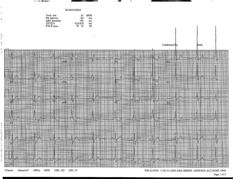
Shown below is an electrocardiogram of Wolff-Parkinson-White syndrome.

Shown below is an electrocardiogram of Wolff-Parkinson-White syndrome.

Shown below is an electrocardiogram of Wolff-Parkinson-White syndrome (antero-lateral pathway).

Shown below is an electrocardiogram of Wolff-Parkinson-White syndrome (antero-septal pathway).

Shown below is an electrocardiogram of Wolff-Parkinson-White syndrome (antero-septal pathway).

Shown below is an electrocardiogram of Wolff-Parkinson-White syndrome (epicardial pathway).

-
Wolf Parkinson White Left Posterior Pathway
-
Wolf Parkinson White Syndrome Posteroseptal Pathway

Shown below is an EKG showing abnormal QRS form with delta waves seen best in the V leads.

-
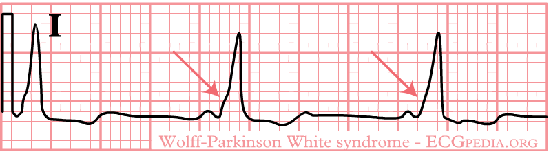
The upstroke of the QRS-complex is 'slurred', resulting in a delta-wave (arrow).
-
Delta waves in a patient with Wolff-Parkinson-White Syndrome (WPW)
-
WPW on a 12 lead ECG.
-
Another example of WPW on a 12 lead ECG.
-
12 lead EKG: Wolff Parkinson White Syndrome
-
12 lead EKG: Wolff Parkinson White Syndrome Type I. Courtesy of Dr Jose Ganseman
-
12 lead EKG: Wolff Parkinson White Syndrome Type I. Courtesy of Dr Jose Ganseman
-
12 lead EKG: Wolff Parkinson White Syndrome Type I. Courtesy of Dr Jose Ganseman
-
12 lead EKG: Wolff Parkinson White Syndrome Type II. Courtesy of Dr Jose Ganseman
-
12 lead EKG: Wolff Parkinson White Syndrome Type II. Courtesy of Dr Jose Ganseman
-
12 lead EKG: Wolff Parkinson White Syndrome Type II. Courtesy of Dr Jose Ganseman
-
12 lead EKG: Wolff Parkinson White Syndrome Type II. Courtesy of Dr Jose Ganseman
-
12 lead EKG: Wolff Parkinson White Syndrome Type II. Courtesy of Dr Jose Ganseman
-
WPW syndrome with an orthodromic circus movement tachycardia: Narrow complex tachycardia with a rate of 200 bpm (RR interval 320 ms). After 5 cycles, the tachycardia suddenly stops and four multiform complexes are seen without any P waves. These complexes should be regarded as a polymorphic ventricular tachycardia, which is not uncommon after an adenosine-terminated supraventricular tachycardia. A 5th complex is preceded by a P wave. The subsequent 4 complexes show a widened QRS complex and all are immediately preceded by a P wave. The initial phase of the QRS complex is slurred and positive in all available leads. Sinus rhythm continues thereafter with gradual abbreviation of the QRS complex until a 120 msec wide QRS complex remains.
-
The same patient's EKG during sinus rhythm. A discrete Δ wave is clearly visible. The morphology of the Δ wave suggests a left posterior Kent bundle.
Sources
Copyleft images obtained courtesy of ECGpedia, http://en.ecgpedia.org/index.php?title=Special:NewFiles&offset=&limit=500