Thin layer chromatography
Template:Infobox chemical analysis
Thin layer chromatography (TLC) is a chromatography technique used to separate chemical compounds.[1] It involves a stationary phase consisting of a thin layer of adsorbent material, usually silica gel, aluminium oxide, or cellulose immobilized onto a flat, inert carrier sheet. A liquid phase consisting of the solution to be separated is then dissolved in an appropriate solvent and is drawn up the plate via capillary action, separating the experimental solution based on the polarity of the components of the compound in question.
Its wide range of uses include
- assaying radiochemical purity of radiopharmaceuticals
- determination of the pigments a plant contains
- detection of pesticides or insecticides in food
- analysing the dye composition of fibers in forensics, or
- identifying compounds present in a given substance
It is a quick, generic method for organic reaction monitoring.
Plate preparation
TLC plates are made by mixing the adsorbent, such as silica gel, with a small amount of inert binder like calcium sulfate (gypsum) and water. This mixture is spread as a thick slurry on an unreactive carrier sheet, usually glass, thick aluminum foil, or plastic, and the resultant plate is dried and activated by heating in an oven for thirty minutes at 110 °C. The thickness of the adsorbent layer is typically around 0.1–0.25 mm for analytical purposes and around 1–2 mm for preparative TLC. Every type of chromatography contains a mobile phase and a stationary phase.
Technique
The process is similar to paper chromatography with the advantage of faster runs, better separations, and the choice between different stationary phases. Because of its simplicity and speed TLC is often used for monitoring chemical reactions and for the qualitative analysis of reaction products.
A small spot of solution containing the sample is applied to a plate, about one centimeter from the base. The plate is then dipped in to a suitable solvent, such as ethanol or water, and placed in a sealed container. The solvent moves up the plate by capillary action and meets the sample mixture, which is dissolved and is carried up the plate by the solvent. Different compounds in the sample mixture travel at different rates owing to differences in solubility in the solvent, and owing to differences in their attraction to the stationary phase
Separation of compounds is based on the competition of the solute and the mobile phase for binding places on the stationary phase. For instance, if normal phase silica gel is used as the stationary phase it can be considered polar. Given two compounds which differ in polarity, the most polar compound has a stronger interaction with the silica and is therefore more capable to dispel the mobile phase from the binding places. Consequently, the less polar compound moves higher up the plate (resulting in a higher Rf value). If the mobile phase is changed to a more polar solvent or mixture of solvents, it is more capable of dispelling solutes from the silica binding places and all compounds on the TLC plate will move higher up the plate. Practically this means that if you use a mixture of ethyl acetate and heptane as the mobile phase, adding more ethyl acetate results in higher Rf values for all compounds on the TLC plate. Changing the polarity of the mobile phase will not result in reversed order of running of the compounds on the TLC plate. If a reversed order of running of the compounds is desired, an apolar stationary phase should be used, such as C18-functionalized silica.
The appropriate solvent in context of Thin layer chromatography will be one which differs from the stationary phase material in polarity. If polar solvent is used to dissolve the sample and spot is applied over polar stationary phase TLC, the sample spot will grow radially due to capillary action, which is not advisable as one spot may mix with the other. Hence, to restrict the radial growth of sample-spot, the solvent used for dissolving samples in order to apply them on plates should be as non-polar or semi-polar as possible when the stationary phase is polar, and vice-versa.
Analysis
As the chemicals being separated may be colorless, several methods exist to visualize the spots:
- Often a small amount of a fluorescent compound, usually manganese-activated zinc silicate, is added to the adsorbent that allows the visualization of spots under a blacklight (UV254). The adsorbent layer will thus fluoresce light green by itself, but spots of analyte quench this fluorescence.
- Iodine vapors are a general unspecific color reagent
- Specific color reagents exist into which the TLC plate is dipped or which are sprayed onto the plate
Once visible, the Rf value , or Retention factor, of each spot can be determined by dividing the distance traveled by the product by the total distance traveled by the solvent (the solvent front). These values depend on the solvent used, and the type of TLC plate, and are not physical constants.
Applications
In organic chemistry, reactions are qualitatively monitored with TLC. Spots sampled with a capillary tube are placed on the plate: a spot of starting material, a spot from the reaction mixture, and a "co-spot" with both. A small (3 by 7 cm) TLC plate takes a couple of minutes to run. The analysis is qualitative, and it will show if starting material has disappeared, product has appeared, and how many products are generated. Unfortunately, TLC's from low-temperature reactions may give misleading results, because the sample is warmed to room temperature in the capillary. One such reaction is DIBALH reduction of ester to aldehyde.
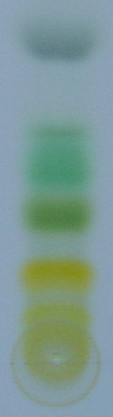
As an example the chromatography of an extract of green leaves (for example spinach) in 7 stages of development. Carotene elutes quickly and is only visible until step 2. Chlorophyll A and B are halfway in the final step and lutein the first compound staining yellow.
-
Step 1
-
Step 2
-
Step 3
-
Step 4
-
Step 5
-
Step 6
-
 Step 7
In one study TLC has been applied in the screening of organic reactions[2] for example in the fine-tuning of BINAP synthesis from 2-naphtol. In this method the alcohol and catalyst solution (for instance iron(III) chloride) are place separately on the base line, then reacted and then instantly analyzed.
References
- ↑ Vogel's Textbook of Practical Organic Chemistry (5th Edition) (Hardcover) by A.I. Vogel (Author), A.R. Tatchell (Author), B.S. Furnis (Author), A.J. Hannaford (Author), P.W.G. Smith ISBN 0582462363
- ↑ TLC plates as a convenient platform for solvent-free reactions Jonathan M. Stoddard, Lien Nguyen, Hector Mata-Chavez and Kelly Nguyen Chem. Commun., 2007, 1240 - 1241, doi:10.1039/b616311d
- Hand book of Thin Layer Chromatography,Sherma, J.and Fried, B. (authors) 3rd ed. Marcel Dekker, New York.
See also
ar:كروموتغرافيا الطبقة الرقيقة
bs:Tankoslojna hromatografija
ca:Cromatografia de capa fina
cs:Chromatografie na tenké vrstvě
de:Dünnschichtchromatografie
it:Cromatografia su strato sottile
nl:Dunnelaagchromatografie
no:Tynnsjiktkromatografi
fi:Ohutkerroskromatografia
sv:Tunnskiktskromatografi