Uploads by Nawal Muazam
Jump to navigation
Jump to search
This special page shows all uploaded files.
| Date | Name | Thumbnail | Size | Description | Versions |
|---|---|---|---|---|---|
| 23:13, 14 December 2015 | Myelodysplastic syndrome with osteomyelosclerosis.jpg (file) | 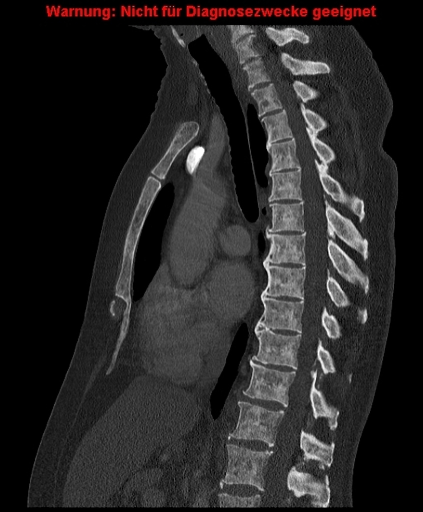 |
311 KB | Case courtesy of Dr Björn Jobke, <a href="http://radiopaedia.org/">Radiopaedia.org</a>. From the case <a href="http://radiopaedia.org/cases/40566">rID: 40566</a> | 1 |
| 16:37, 2 December 2015 | Mast cell leukemia peripheral blood smear.jpg (file) | 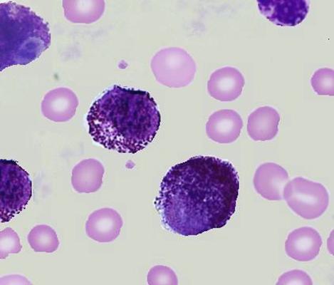 |
18 KB | Peripheral blood showing mast cell leukemia. | 1 |
| 21:37, 1 December 2015 | Mast cell leukemia.jpg (file) | 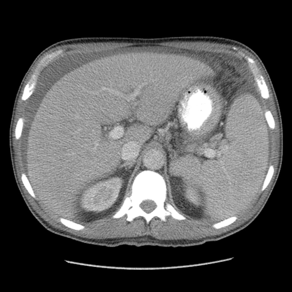 |
99 KB | CT of the abdomen demonstrates ascites, hepatosplenomegaly and upper abdominal lymphadenopathy. Windowing to bone confirms the diffuse sclerosis seen on the plain films. | 1 |
| 21:17, 17 November 2015 | Facial-hemangioma.jpg (file) | 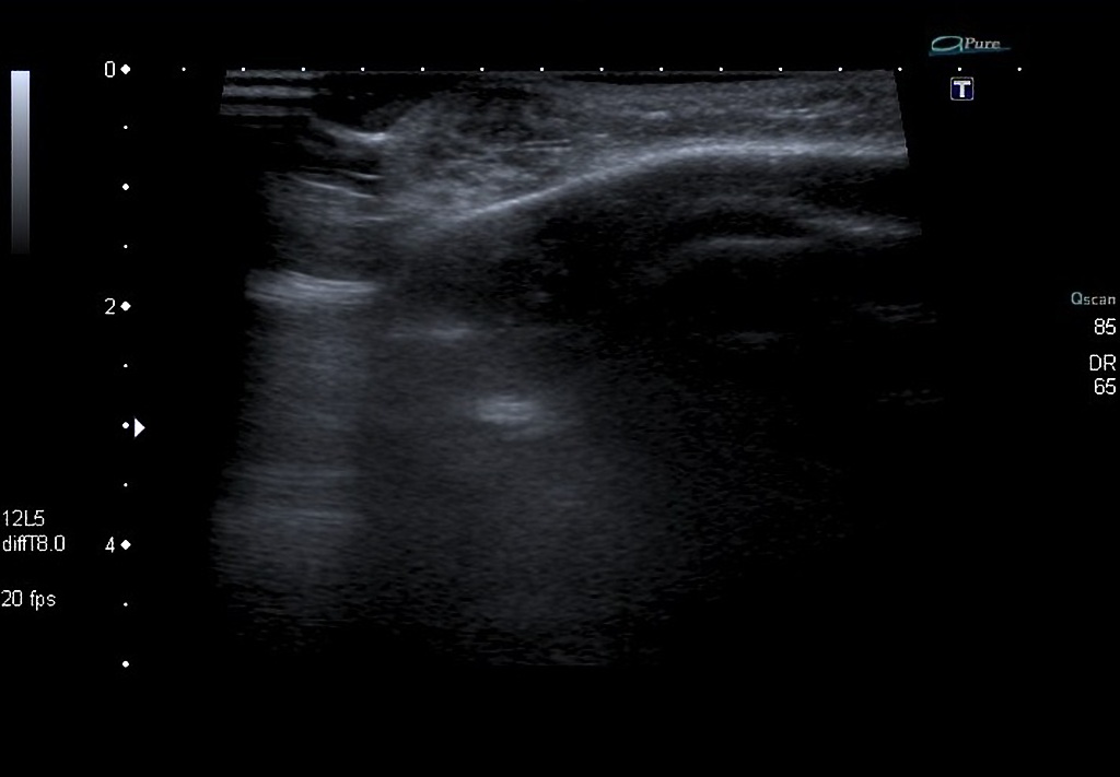 |
77 KB | Color Doppler image demonstrates a 1.5 cm highly vascular mass consistent with a capillary hemangioma located at the superolateral margin of the right orbit. | 1 |
| 22:13, 13 November 2015 | Hepatoblastoma ultrasound.jpg (file) | 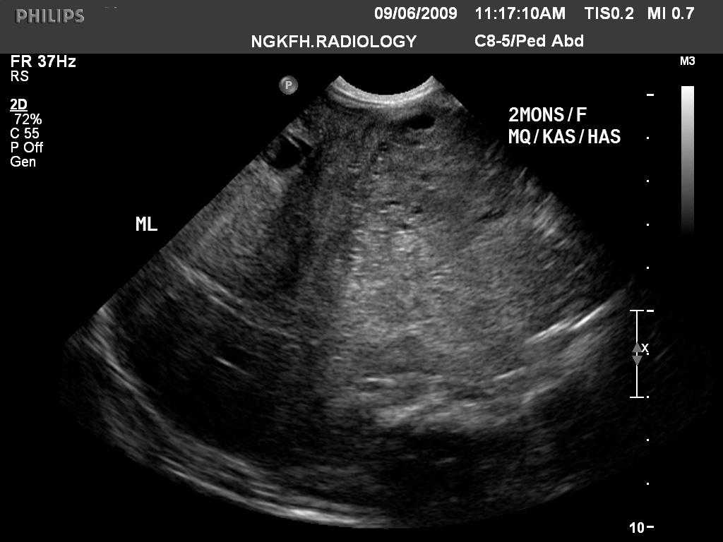 |
88 KB | Ultrasound demonstrates a large heterogeneous, echogenic, and hypervascular mass. | 1 |
| 21:40, 13 November 2015 | Hepatoblastoma CT scan2.jpg (file) | 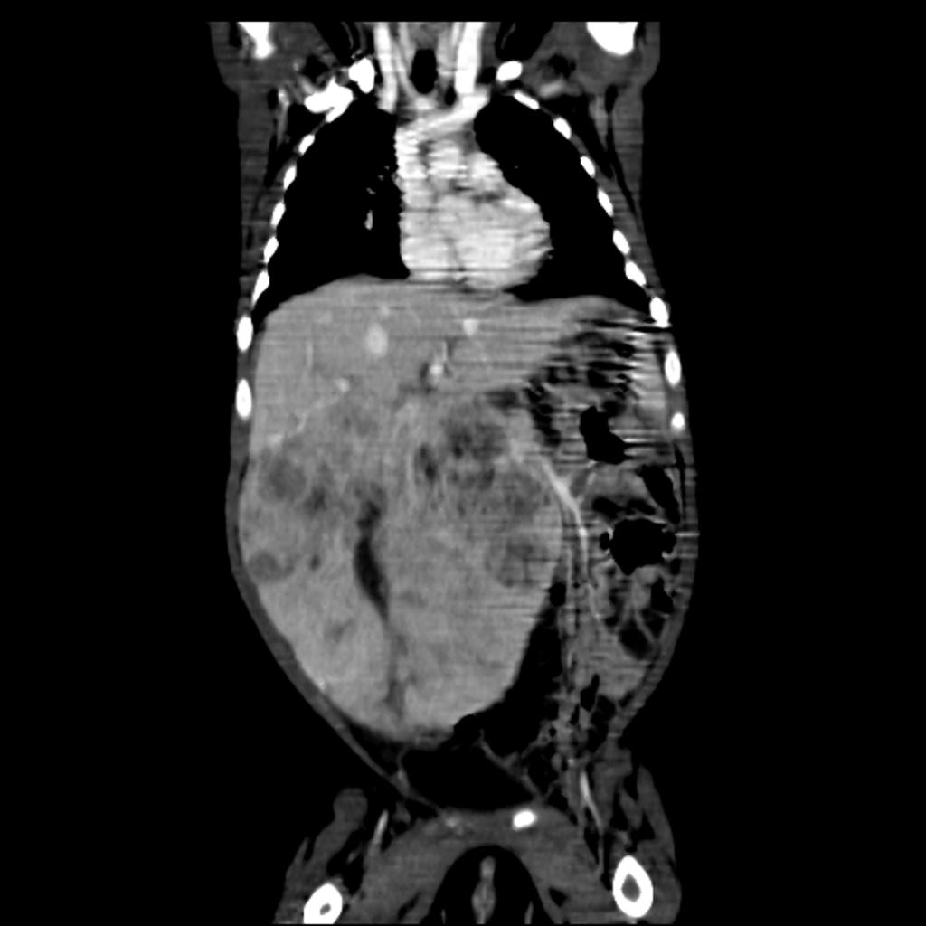 |
57 KB | CT shows a large heterogenous predominantly hypodense mass lesion arising from right lobe of liver with a chunky calcification. | 1 |
| 21:36, 13 November 2015 | Hepatoblastoma CT scan.jpg (file) | 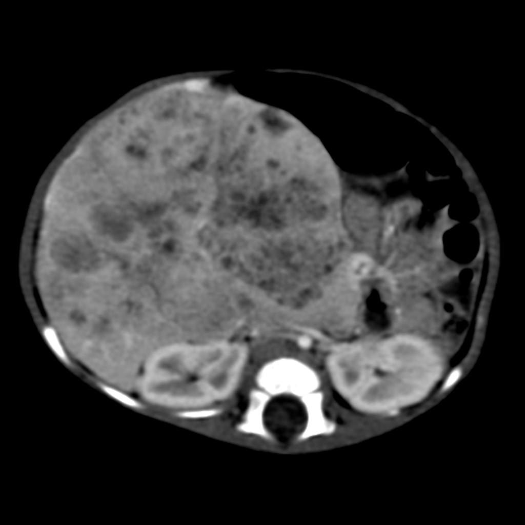 |
47 KB | CT shows a large heterogenous predominantly hypodense mass lesion arising from right lobe of liver with a chunky calcification. | 1 |
| 19:08, 12 November 2015 | Cavernous hemangioma histopathology2.jpg (file) | 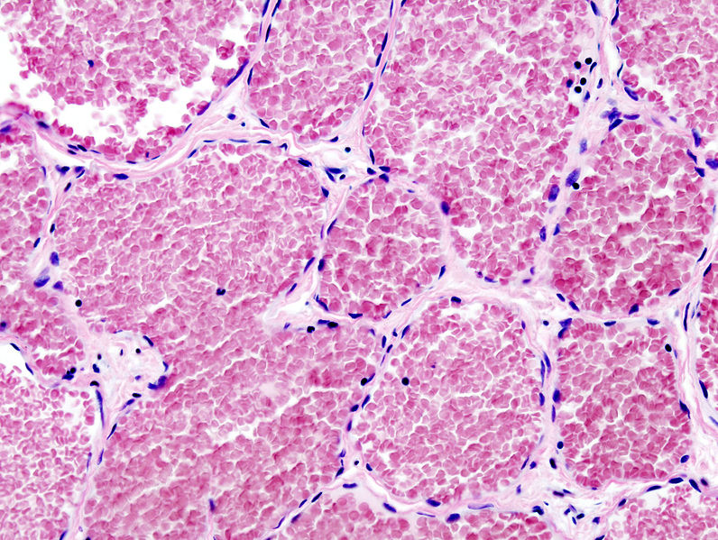 |
198 KB | Histopathological image reprsenting a cavernous hemangioma of the liver. | 1 |
| 18:55, 12 November 2015 | Cavernous hemangioma histopathology.jpg (file) | 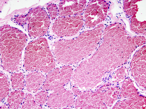 |
114 KB | Histopathological image representing a cavernous hemangioma of the liver. Surgical excision of the lesion for the impending risk for rupture. H&E stain. | 1 |
| 18:48, 12 November 2015 | Capillary hemangioma very high magnification.jpg (file) | 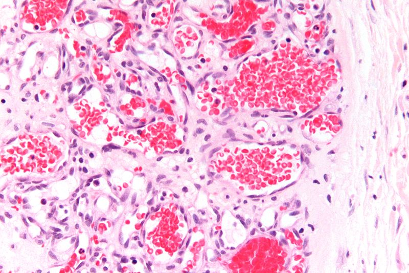 |
106 KB | Very high magnification micrograph of a capillary hemangioma. H&E stain. | 1 |
| 18:37, 12 November 2015 | Capillary hemangioma intermediate magnification.jpg (file) | 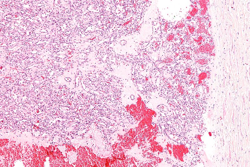 |
176 KB | Intermediate magnification micrograph of a capillary hemangioma. H&E stain. | 1 |
| 21:45, 9 November 2015 | Hepatic hemangioma MRI.jpg (file) | 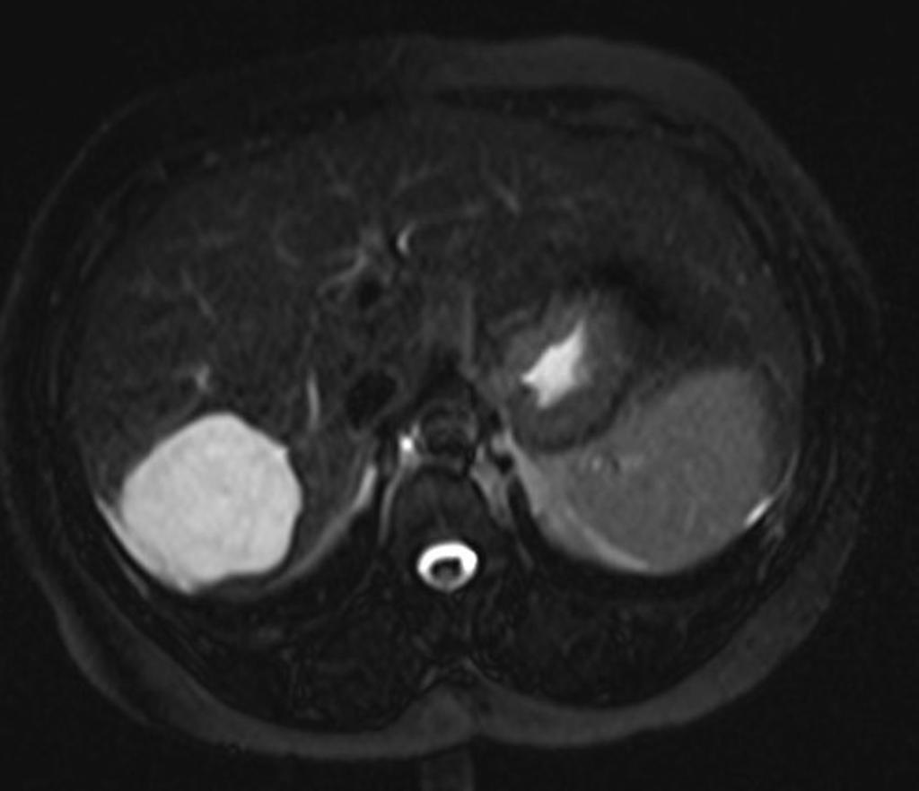 |
46 KB | MRI reveals a lobullated T2 hyperintense lesion in the right lobe segments VI and VII. | 1 |
| 18:18, 7 November 2015 | Hepatocellular adenoma high magnification.jpg (file) | 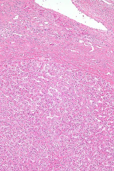 |
114 KB | High magnification micrograph of a hepatic adenoma. H&E stain. | 1 |
| 18:06, 7 November 2015 | Hepatic adenoma low magnification.jpg (file) | 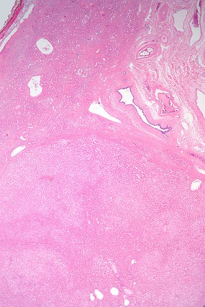 |
71 KB | Low magnification micrograph of a hepatic adenoma. H&E stain. | 1 |
| 17:46, 7 November 2015 | Hepatoblastoma high magnification 2.jpg (file) | 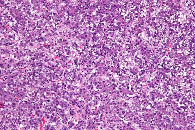 |
191 KB | High magnification micrograph of a hepatoblastoma, a type of liver cancer found in infants and young children. H&E stain. | 1 |
| 17:34, 7 November 2015 | Hepatoblastoma high magnification.jpg (file) | 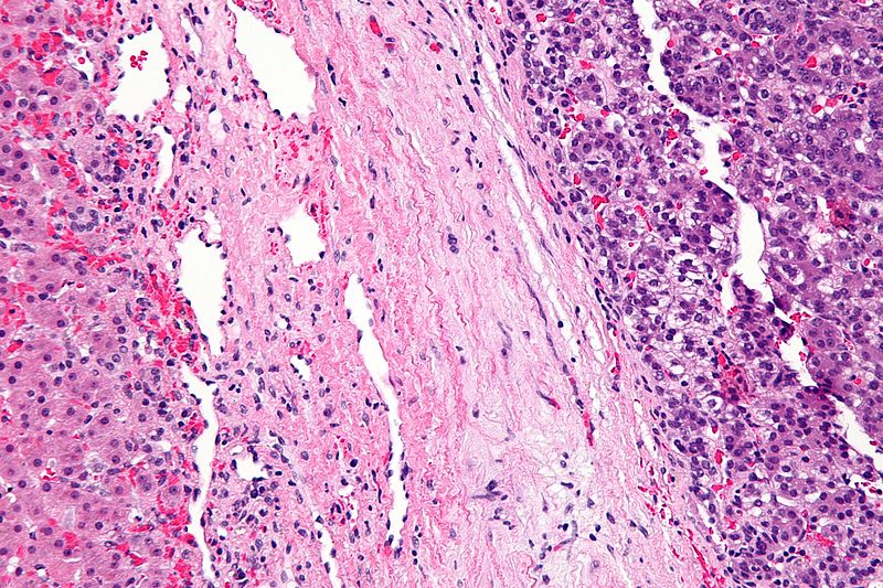 |
181 KB | High magnification micrograph of a hepatoblastoma, a type of liver cancer found in infants and young children. H&E stain. | 1 |
| 17:13, 7 November 2015 | Cavernous liver hemangioma high magnification.jpg (file) | 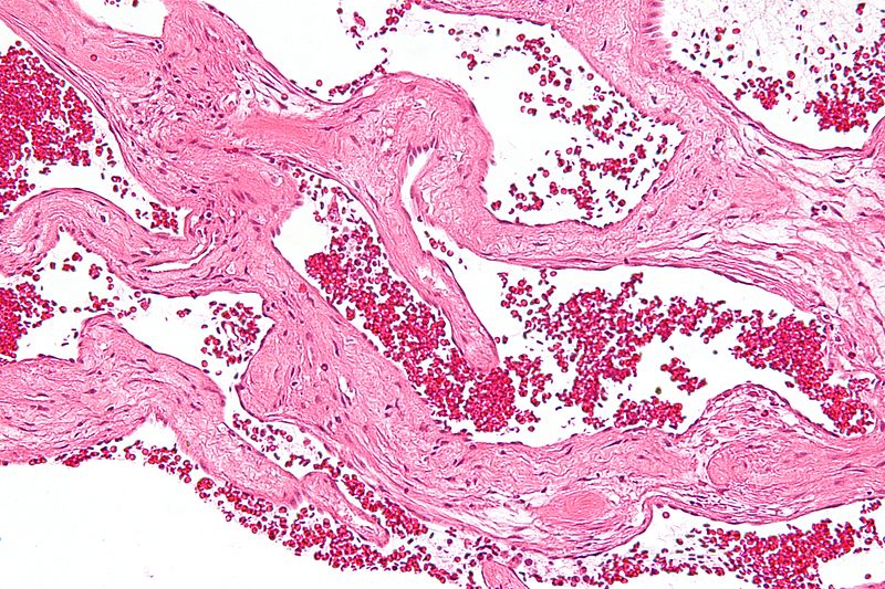 |
178 KB | English: High magnification micrograph of a cavernous hemangioma of the liver, also hepatic cavernous hemangioma, liver hemangioma, cavernous liver hemangioma. H&E stain. No liver tissue is seen. | 1 |
| 17:02, 3 November 2015 | Hepatoblastoma microscopy1.jpg (file) | 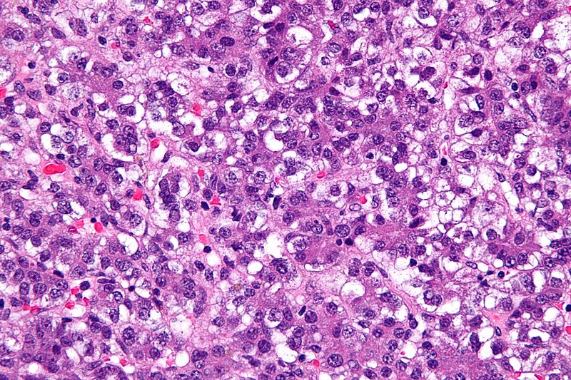 |
182 KB | Very high magnification micrograph of a hepatoblastoma, a type of liver cancer found in infants and young children. H&E stain. | 1 |
| 15:19, 27 October 2015 | Giant-hepatic-haemangiomata.png (file) | 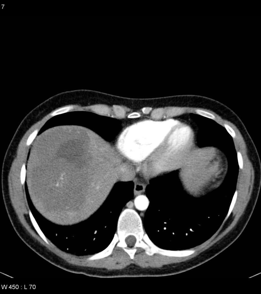 |
303 KB | 1 | |
| 15:01, 27 October 2015 | Giant-hepatic-haemangiomata (2).jpg (file) | 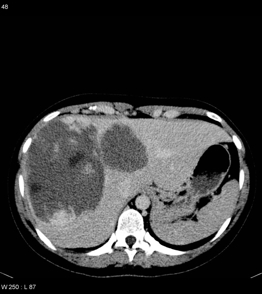 |
82 KB | Case courtesy of Dr Natalie Yang, <a href="http://radiopaedia.org/">Radiopaedia.org</a>. From the case <a href="http://radiopaedia.org/cases/7014">rID: 7014</a> | 1 |
| 14:54, 27 October 2015 | Giant-hepatic-haemangiomata.jpg (file) | 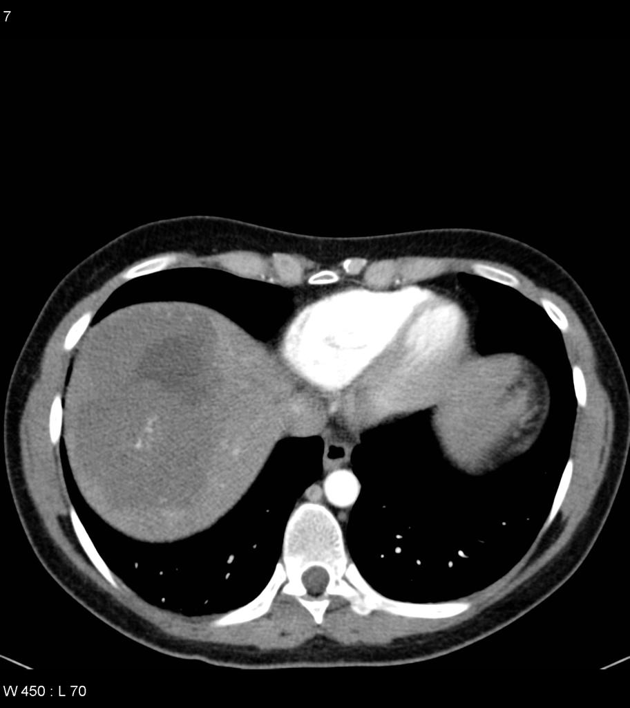 |
62 KB | Case courtesy of Dr Natalie Yang, <a href="http://radiopaedia.org/">Radiopaedia.org</a>. From the case <a href="http://radiopaedia.org/cases/7014">rID: 7014</a> | 1 |
| 19:51, 20 October 2015 | Cavernous liver hemangioma - intermed mag.jpg (file) | 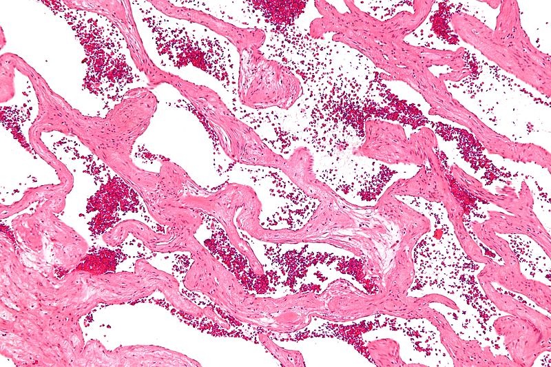 |
177 KB | 2 | |
| 15:53, 16 October 2015 | Hepatic-adenoma-T2 fat sat.jpg (file) | 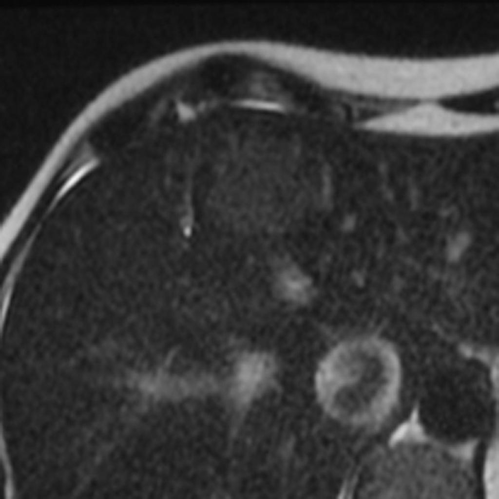 |
55 KB | Case courtesy of Dr Natalie Yang, <a href="http://radiopaedia.org/">Radiopaedia.org</a>. From the case <a href="http://radiopaedia.org/cases/6953">rID: 6953</a> | 1 |
| 15:47, 16 October 2015 | Axial T1 out of phase hepatic adenoma.jpg (file) | 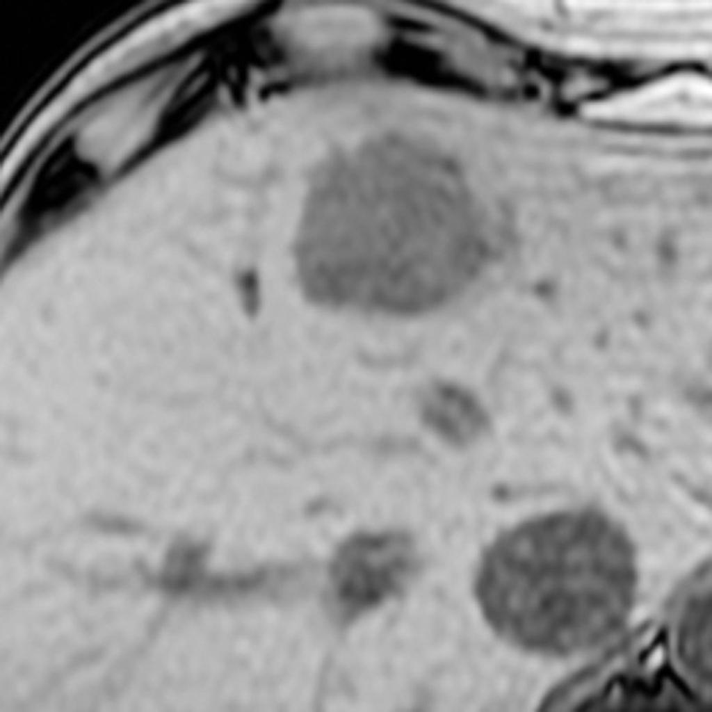 |
55 KB | 1 | |
| 19:14, 15 October 2015 | Hepatic adenoma11.jpg (file) | 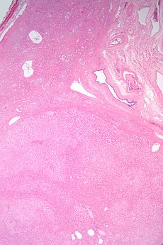 |
20 KB | 2 |