Renal pyramids
|
WikiDoc Resources for Renal pyramids |
|
Articles |
|---|
|
Most recent articles on Renal pyramids Most cited articles on Renal pyramids |
|
Media |
|
Powerpoint slides on Renal pyramids |
|
Evidence Based Medicine |
|
Clinical Trials |
|
Ongoing Trials on Renal pyramids at Clinical Trials.gov Trial results on Renal pyramids Clinical Trials on Renal pyramids at Google
|
|
Guidelines / Policies / Govt |
|
US National Guidelines Clearinghouse on Renal pyramids NICE Guidance on Renal pyramids
|
|
Books |
|
News |
|
Commentary |
|
Definitions |
|
Patient Resources / Community |
|
Patient resources on Renal pyramids Discussion groups on Renal pyramids Patient Handouts on Renal pyramids Directions to Hospitals Treating Renal pyramids Risk calculators and risk factors for Renal pyramids
|
|
Healthcare Provider Resources |
|
Causes & Risk Factors for Renal pyramids |
|
Continuing Medical Education (CME) |
|
International |
|
|
|
Business |
|
Experimental / Informatics |
Editor-In-Chief: C. Michael Gibson, M.S., M.D. [1]
Renal pyramids (or malpighian pyramids) are cone-shaped tissues of the kidney. The renal medulla is made up of 8 to 18 of these conical subdivisions. The broad base of each pyramid faces the renal cortex, and its apex, or papilla, points internally. The pyramids appear striped because they are formed by straight parallel segments of nephrons.
Additional images
-
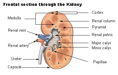 Frontal section through the kidney

The base of each pyramid originates at the corticomedullary border and the apex terminates in a papilla, which lies within a minor calyx.