Renal medulla
|
WikiDoc Resources for Renal medulla |
|
Articles |
|---|
|
Most recent articles on Renal medulla Most cited articles on Renal medulla |
|
Media |
|
Powerpoint slides on Renal medulla |
|
Evidence Based Medicine |
|
Clinical Trials |
|
Ongoing Trials on Renal medulla at Clinical Trials.gov Trial results on Renal medulla Clinical Trials on Renal medulla at Google
|
|
Guidelines / Policies / Govt |
|
US National Guidelines Clearinghouse on Renal medulla NICE Guidance on Renal medulla
|
|
Books |
|
News |
|
Commentary |
|
Definitions |
|
Patient Resources / Community |
|
Patient resources on Renal medulla Discussion groups on Renal medulla Patient Handouts on Renal medulla Directions to Hospitals Treating Renal medulla Risk calculators and risk factors for Renal medulla
|
|
Healthcare Provider Resources |
|
Causes & Risk Factors for Renal medulla |
|
Continuing Medical Education (CME) |
|
International |
|
|
|
Business |
|
Experimental / Informatics |
Overview
The renal medulla is the innermost part of the kidney. The renal medulla is split up into a number of sections, known as the renal pyramids. Blood enters into the kidney via the renal artery, which then splits up to form the arcuate arterioles. The arcuate arterioles each in turn branch into interlobar arterioles, which finally reach the glomeruli. At the glomerulus the blood reaches a highly disfavourable pressure gradient and a large exchange surface area, which forces the serum portion of the blood out of the vessel into the renal tubules. Flow continues through the renal tubules, including the proximal tubule, the Loop of Henle, and finally leaves the kidney by means of the collecting duct, leading to the renal urethra.
Additional images
-
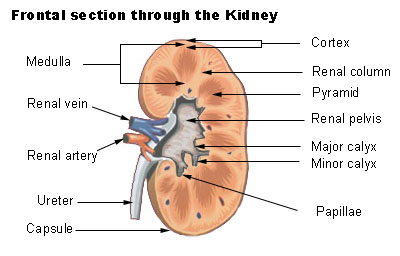 Frontal section through the kidney
 -
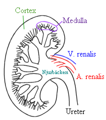 Kidney


External links
Template:Kidney Template:Anatomy-stub Template:WH Template:WS