Olfactory bulb
|
WikiDoc Resources for Olfactory bulb |
|
Articles |
|---|
|
Most recent articles on Olfactory bulb Most cited articles on Olfactory bulb |
|
Media |
|
Powerpoint slides on Olfactory bulb |
|
Evidence Based Medicine |
|
Clinical Trials |
|
Ongoing Trials on Olfactory bulb at Clinical Trials.gov Trial results on Olfactory bulb Clinical Trials on Olfactory bulb at Google
|
|
Guidelines / Policies / Govt |
|
US National Guidelines Clearinghouse on Olfactory bulb NICE Guidance on Olfactory bulb
|
|
Books |
|
News |
|
Commentary |
|
Definitions |
|
Patient Resources / Community |
|
Patient resources on Olfactory bulb Discussion groups on Olfactory bulb Patient Handouts on Olfactory bulb Directions to Hospitals Treating Olfactory bulb Risk calculators and risk factors for Olfactory bulb
|
|
Healthcare Provider Resources |
|
Causes & Risk Factors for Olfactory bulb |
|
Continuing Medical Education (CME) |
|
International |
|
|
|
Business |
|
Experimental / Informatics |
Overview
The olfactory bulb is a structure of the vertebrate forebrain involved in olfaction, the perception of odors.
Anatomy
In most vertebrates, the olfactory bulb is the most rostral (forward) part of the brain. In humans, however, the olfactory bulb is on the inferior (bottom) side of the brain. The olfactory bulb is supported and protected by the cribriform plate which in mammals, separates it from the olfactory epithelium, and which is perforated by olfactory nerve axons. The bulb is divided into two distinct structures, the main olfactory bulb, and the accessory olfactory bulb.
Main olfactory bulb
The main olfactory bulb has a multi-layered cellular architecture. In order from the surface to the center of the bulb the layers are
- Glomerular layer
- External plexiform layer
- Mitral cell layer
- Internal plexiform layer
- Granule cell layer
The glomerular layer receives direct input from olfactory nerves, made up of the axons from approximately ten million olfactory receptor neurons in the olfactory mucosa, a region of the nasal cavity. The ends of the axons cluster in spherical structures known as glomeruli such that each glomerulus receives input primarily from olfactory receptor neurons that express the same olfactory receptor. Glomeruli are also permeated by dendrites from neurons called mitral cells, which in turn output to the olfactory cortex. Numerous interneuron types exist in the olfactory bulb including periglomerular cells which synapse within and between glomeruli, and granule cells which synapse with mitral cells.
Accessory olfactory bulb
The accessory olfactory bulb, which resides on the dorsal-posterior region of the main olfactory bulb, forms a parallel pathway independent from the main olfactory bulb. It is the second processing stage of the accessory olfactory system. It receives axonal input from the vomeronasal organ, a distinct sensory epithelium from the main olfactory epithelium that detects pheremones, among other chemical stimuli. Like the main olfactory bulb, axonal input to the accessory olfactory bulb forms synapses with mitral cells within glomeruli. However, mitral cells in the accessory olfactory bulb project their axons to targets in the amygdala and hypothalamus where they may influence aggressive and mating behavior.
Function

The olfactory bulb transmits smell information from the nose to the brain, and is thus necessary for a proper sense of smell. As a neural circuit, the olfactory bulb has one source of sensory input (axons from olfactory receptor neurons of the olfactory epithelium), and one output (mitral cell axons). As a result, it is generally assumed that it functions as a filter, as opposed to an associative circuit that has many inputs and many outputs. However, the olfactory bulb also receives "top-down" information from such brain areas as the amygdala, neocortex, hippocampus, locus coeruleus, and substantia nigra. With this in mind, its potential functions can be placed into four non-exclusive categories:
- enhancing discrimination between odors.
- enhancing sensitivity of odor detection.
- filtering out many background odors to enhance the transmission of a few select odors.
- permitting higher brain areas involved in arousal and attention to modify the detection or the discrimination of odors.
While all of these functions could theoretically arise from the olfactory bulb's circuit layout, it is unclear which, if any, of these functions are performed exclusively by the olfactory bulb. By analogy to similar parts of the brain such as the retina, many researchers have focused on how the olfactory bulb filters incoming information from receptor neurons in space, or how it filters incoming information in time. At the core of these proposed filters are the two classes of interneurons; the periglomerular cells, and the granule cells.
Mitral cells are connected by interneurons known as granule cells, which by some theories produce lateral inhibition between mitral cells. It is not clear what the functional role of lateral inhibition would be, though it may be involved in boosting the signal-to-noise ratio of odor signals by silencing the basal firing rate of surrounding non-activated neurons. The synapse between mitral and granule cells is of a rare class of synapses that are "dendro-dendritic" which means that both sides of the synapse are dendrites that release neurotransmitter. In this specific case, mitral cells release the excitatory neurotransmitter glutamate, and granule cells release the inhibitory neurotransmitter Gamma-aminobutyric acid (GABA). As a result of its bi-directionality, the dendro-dendritic synapse can cause mitral cells to inhibit themselves (auto-inhibition), as well as neighboring mitral cells (lateral inhibition).
Evolution
Comparing the structure of the olfactory bulb between species such as the fruit fly (where it is called the antennal lobe), the Leopard Frog and the lab mouse reveals that they all share the same fundamental layout (five layers containing the nuclei of three major cell types, see "Anatomy" for details), despite being dissimilar in shape and size. It is quite possible, however, that function has not been conserved with structure, indicating that olfactory bulbs of distant species may be paralogs of each other. An intriguing alternative possibility states olfactory bulb structure is conserved because it contains an optimal solution to a computational problem experienced by all olfactory systems, and thus may have evolved independently in different species.
References
- Shepherd, G. The Synaptic Organization of the Brain, Oxford University Press, 5th edition (November, 2003). ISBN 0-19-515956-X
- Halpern M, Martinez-Marcos A, Structure and function of the vomeronasal system: an update. Progress in Neurobiology; 70(245-318), 2003.
- Ache BW, Young JM, Olfaction: Diverse Species, Conserved Principles. Neuron; 48(417-430), 2005.
Additional images
-
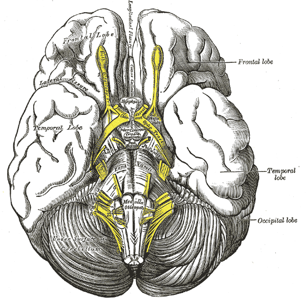
Base of brain.
-
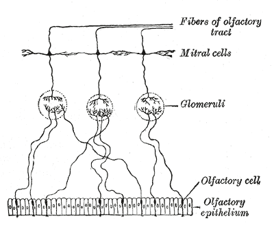
Plan of olfactory neurons.
-
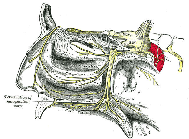
The pterygopalatine ganglion and its branches.


External links
Template:Olfactory system Template:Prosencephalon
de:Riechkolben fa:پیاز بویایی id:Bulbus olfaktorius la:Bulbus olfactorius nl:Bulbus olfactorius Template:WH