Meningioma classification

Jump to navigation Jump to search

Meningioma Microchapters

Home

Patient Information

Overview

Historical Perspective

Classification

Pathophysiology

Causes

Differentiating Meningioma from other Diseases

Epidemiology and Demographics

Risk Factors

Screening

Natural History, Complications and Prognosis

Diagnosis

Diagnostic Study of Choice

History and Symptoms

Physical Examination

Laboratory Findings

Electrocardiogram

X-ray

Echocardiography and Ultrasound

CT scan

MRI

Other Imaging Findings

Other Diagnostic Studies

Treatment

Medical Therapy

Interventions

Surgery

Primary Prevention

Secondary Prevention

Cost-Effectiveness of Therapy

Future or Investigational Therapies

Case Studies

Case #1

Meningioma classification On the Web

Most recent articles

Most cited articles

Review articles

CME Programs

Powerpoint slides

Images

American Roentgen Ray Society Images of Meningioma classification

All Images
X-rays
Echo and Ultrasound
CT Images
MRI

Ongoing Trials at Clinical Trials.gov

US National Guidelines Clearinghouse

NICE Guidance

FDA on Meningioma classification

CDC on Meningioma classification

Meningioma classification in the news

Blogs on Meningioma classification

Directions to Hospitals Treating Meningioma

Risk calculators and risk factors for Meningioma classification

Editor-In-Chief: C. Michael Gibson, M.S., M.D. [1] Associate Editor(s)-in-Chief: Ifeoma Odukwe, M.D. [2] Haytham Allaham, M.D. [3]

Overview

Meningioma has been classified into 3 groups by the WHO: benign classic meningioma (WHO grade 1), atypical meningioma (WHO grade 2), and anaplastic malignant meningioma (WHO grade 3). The WHO grade 1 group grow very slowly and consists of about 9 variants. About 3 variants correspond to WHO grade II, their association with malignancy is not clear but they grow faster than benign meningiomas. About 2 variants correspond to WHO grade III.

Classification

  • The table below lists the subtypes of meningioma according to the WHO grades:[1][2]
WHO Grade Subtypes

Benign classic meningioma
(WHO grade 1)

  • Meningothelial, fibrous, transitional, psammomatous,
    angiomatous, micrcystic, secretory, lymphoplasmacyte-rich, and metaplastic meningioma
  • They are low-grade tumors that grow very slowly and rarely invade the surrounding tissues

Atypical meningioma
(WHO grade 2)

  • Chordoid, clear cell, and atypical meningioma.
  • These are not clearly malignant, but grow faster than benign meningiomas
  • 4 or more mitotic cells per hpf and/or 3 or more of the following features: small cells, increased cellularity, necrosis, prominent nucleoli, sheeting, &/or brain invasion in an otherwise Grade I tumor

Anaplastic malignant meningioma
(WHO grade 3)

  • Rhabdoid, papillary, and anaplastic meningioma
  • 20 or more mitoses per 10 hpf and/or possess cytological characteristics of an obvious malignancy such that the tumor cell resembles carcinoma, sarcoma, or melanoma.

Gallery

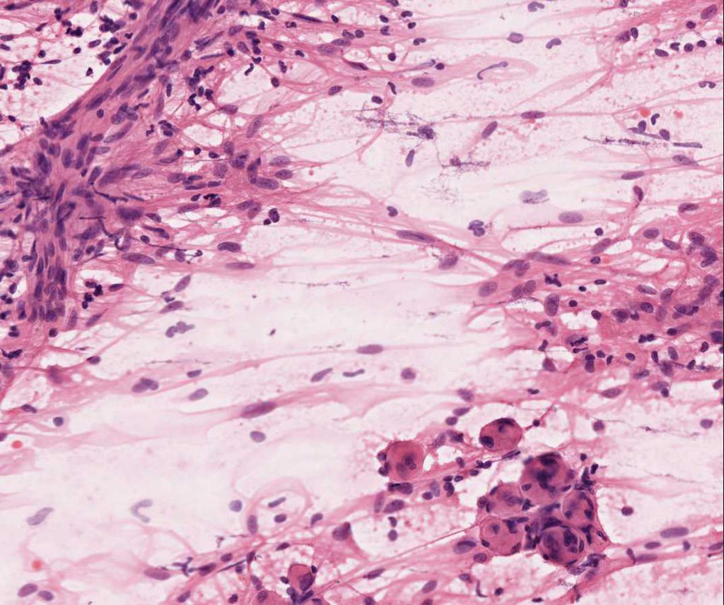
Meningothelial meningioma Source: Case courtesy of Dr Victor Yang, <a href="https://radiopaedia.org/">Radiopaedia.org</a>.


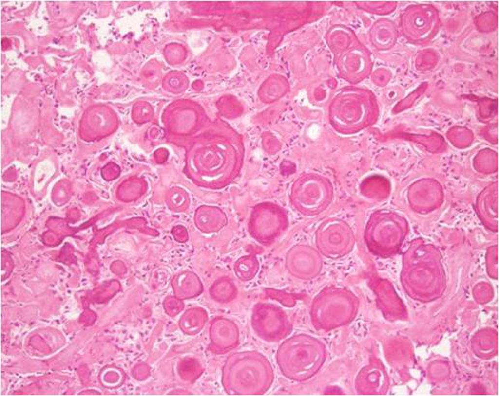
Psammomatous meningioma Source: Case courtesy of A.Prof Frank Gaillard, <a href="https://radiopaedia.org/">Radiopaedia.org</a>.


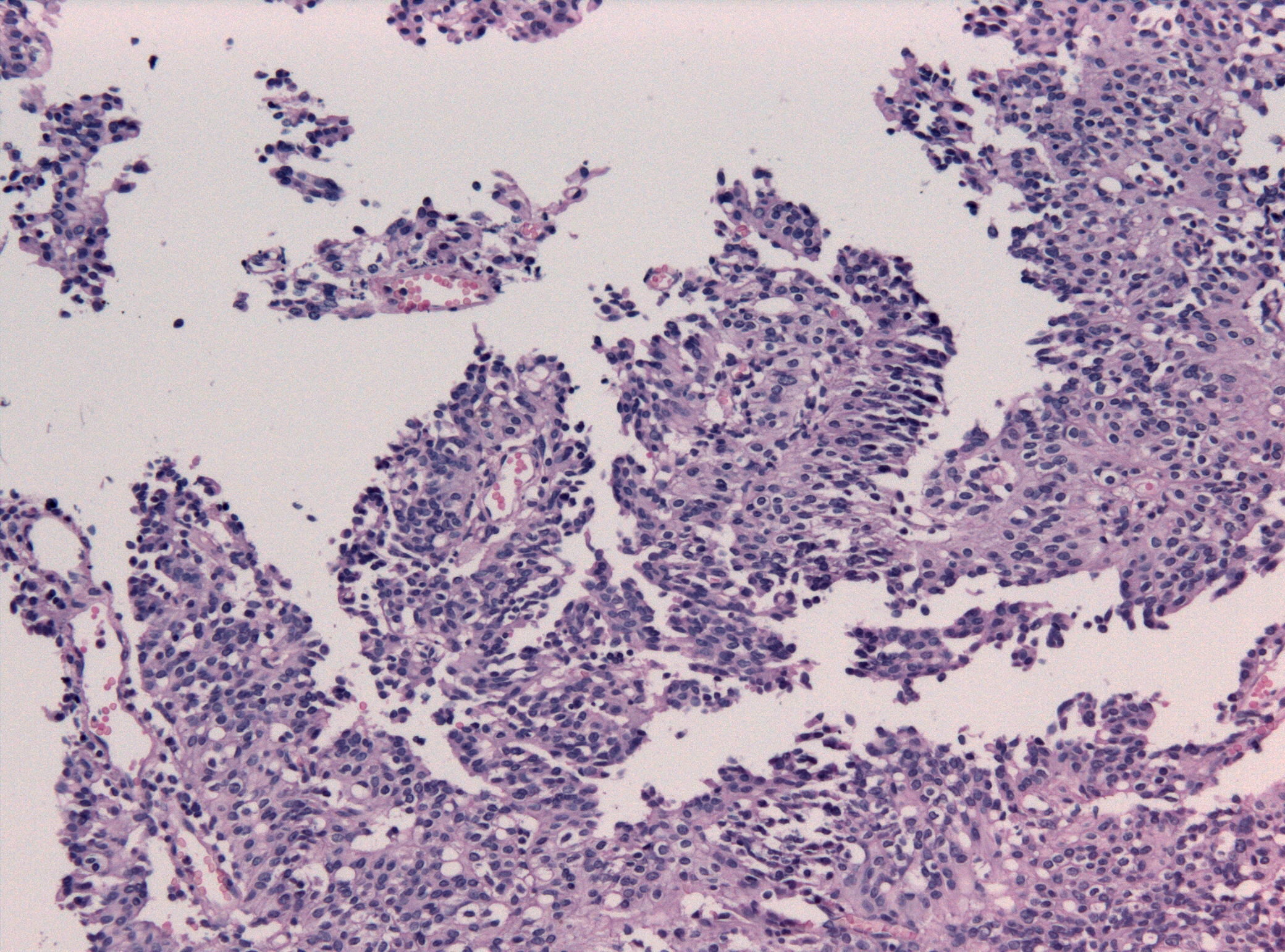
Papillary meningioma Source: Jensflorian-wikimedia commons


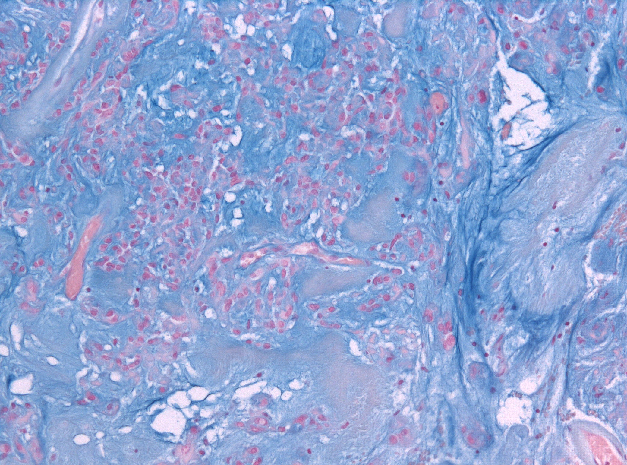
Alcian blue histologic slide showing chordoid meningioma Source: Jensflorian-wikimedia commons


References

  1. 1.0 1.1 Harter, Patrick N.; Braun, Yannick; Plate, Karl H. (2017). "Classification of meningiomas—advances and controversies". Chinese Clinical Oncology. 6 (S1): S2–S2. doi:10.21037/cco.2017.05.02. ISSN 2304-3865.
  2. Commins, Deborah L.; Atkinson, Roscoe D.; Burnett, Margaret E. (2007). "Review of meningioma histopathology". Neurosurgical Focus. 23 (4): E3. doi:10.3171/FOC-07/10/E3. ISSN 1092-0684.