Medial plantar artery
Jump to navigation
Jump to search
Editor-In-Chief: C. Michael Gibson, M.S., M.D. [1]
The medial plantar artery (internal plantar artery), much smaller than the lateral, passes forward along the medial side of the foot.
It is at first situated above the Abductor hallucis, and then between it and the Flexor digitorum brevis, both of which it supplies.
At the base of the first metatarsal bone, where it is much diminished in size, it passes along the medial border of the first toe, anastomosing with the first dorsal metatarsal artery.
Small superficial digital branches accompany the digital branches of the medial plantar nerve and join the plantar metatarsal arteries of the first three spaces.
Additional images
-
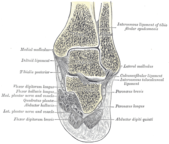 Coronal section through right talocrural and talocalcaneal joints.
