Marburg hemorrhagic fever pathophysiology
|
Marburg hemorrhagic fever Microchapters |
|
Differentiating Marburg hemorrhagic fever from other Diseases |
|---|
|
Diagnosis |
|
Treatment |
|
Case Studies |
|
Marburg hemorrhagic fever pathophysiology On the Web |
|
American Roentgen Ray Society Images of Marburg hemorrhagic fever pathophysiology |
|
Risk calculators and risk factors for Marburg hemorrhagic fever pathophysiology |
Editor-In-Chief: C. Michael Gibson, M.S., M.D. [1]; Associate Editor(s)-in-Chief: Anmol Pitliya, M.B.B.S. M.D.[2]
Overview
Marburg virus is the causative agent of Marburg haemorrhagic fever (MHF). Initial human infection results from prolonged exposure to mines or caves inhabited by Rousettus bat colonies. After the Marburg virus initially transfers from animal host to human, mode of transmission is usually human-to-human and results from direct contact with bodily fluids of infected persons (blood, secretions) other contact fomites contaminated with infectious blood and tissues. Marburg virus primarily infects macrophages and dendritic cells. A cascade of events leads to Hypotension, metabolic disorders, immunosuppression and coagulopathy, finally resulting in multiorgan failure and shock.
Pathophysiology
Pathogen
 |
 |
- Marburg virus is the causative agent of Marburg haemorrhagic fever (MHF). Marburg and Ebola viruses are the two members of the Filoviridae family (filovirus). Though caused by different viruses, the two diseases are clinically similar.
- The viral structure is typical of filoviruses, with long threadlike particles which have a consistent diameter but vary greatly in length from an average of 800 nanometers up to 14,000 nm. Peak infectious activity is at approximately 790 nm.
- Virions contain seven known structural proteins. Four proteins form the nucelocapsid of the Marburg virus: NP, VP35, VP30, and L.[2] While nearly identical to Ebola virus in structure, Marburg virus is antigenically distinct from Ebola virus.
- Marburg virus was the first filovirus to be identified.
Transmission
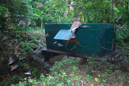
- Initial human infection results from prolonged exposure to mines or caves inhabited by Rousettus bat colonies. The reservoir host of Marburg virus is the African fruit bat, Rousettus aegyptiacus. Marburg virus can infect primates (including humans) and may cause serious disease with high mortality.[3][4]
- After the Marburg virus initially transfers from animal host to human, mode of transmission is usually human-to-human and results from direct contact with bodily fluids of infected persons (blood, secretions) other contact fomites contaminated with infectious blood and tissues.[5]
- Transmission to health-care workers has been reported while treating Marburg patients, mainly due to incorrect or inadequate use of personal protective equipment.
Incubation
- The incubation period (interval from infection to onset of symptoms) varies from 5 to 10 days.
Pathogenesis
Pathogenesis of hemorrhagic fever by Marburg virus is as follows:
- Marburg virus primarily infects macrophages and dendritic cells.[6]
- Infection of dendritic cells leads to paralysis of cellular antiviral response and dysregulation of costimulation of lymphocytes.
- Infection of macrophage leads to the production of proinflammatory mediators including TNF-α, IL-6 and tissue factor.
- TNF-α induce apoptosis in lymphocyte and results in lymphopenia and immunosuppression.
- IL-6 and TNF-α also induces increase in vascular permeability.
- Tissue factor produced by infected macrophages leads to dysregulation of coagulation (e.g., DIC).
- Hepatocyte infection further reinforces coagulation dysregulation due to ineffective production of clotting factors.
- Adrenal cortical cells infection results in hypotension and metabolic disorders.
- Hypotension, metabolic disorders, immunosuppression and coagulopathy results in multiorgan failure and shock.
| Marburg virus | |||||||||||||||||||||||||||||||||||||||||||||||||||||||
| Dendritic cells | Adrenal Cortical cells | Hepatocytes | Macropohages | ||||||||||||||||||||||||||||||||||||||||||||||||||||
| Paralysis of cellular antiviral response | Dysregulated costimulation | ||||||||||||||||||||||||||||||||||||||||||||||||||||||
| Tissue factor | TNF-α IL-6 | TNF-α | |||||||||||||||||||||||||||||||||||||||||||||||||||||
| Endothelial cells | T lymphocytes(CD4+/CD8+ and Natural killer cells | ||||||||||||||||||||||||||||||||||||||||||||||||||||||
| Coagulation dysregulation (DIC) | Increased vascular permeability | TNF-related apoptosis-inducing ligand(TRAIL) | |||||||||||||||||||||||||||||||||||||||||||||||||||||
| Liver dysfunction Inhibition of coagulation factor synthesis | Apoptosis of lymphocytes leading to lymphopenia | ||||||||||||||||||||||||||||||||||||||||||||||||||||||
| Immunosupression | |||||||||||||||||||||||||||||||||||||||||||||||||||||||
| Hypotension Metabolic disorder | Hemorrhagic syndrome | ||||||||||||||||||||||||||||||||||||||||||||||||||||||
| Shock Multiorgan failure | |||||||||||||||||||||||||||||||||||||||||||||||||||||||
References
- ↑ "Electron micrograph (TEM) of the Marburg Hemorrhagic Virus (MHV)".
- ↑ Becker S, Rinne C, Hofsäss U, Klenk HD, Mühlberger E (1998). "Interactions of Marburg virus nucleocapsid proteins". Virology. 249 (2): 406–17. doi:10.1006/viro.1998.9328. PMID 9791031.
- ↑ Adjemian J, Farnon EC, Tschioko F, Wamala JF, Byaruhanga E, Bwire GS, Kansiime E, Kagirita A, Ahimbisibwe S, Katunguka F, Jeffs B, Lutwama JJ, Downing R, Tappero JW, Formenty P, Amman B, Manning C, Towner J, Nichol ST, Rollin PE (2011). "Outbreak of Marburg hemorrhagic fever among miners in Kamwenge and Ibanda Districts, Uganda, 2007". J. Infect. Dis. 204 Suppl 3: S796–9. doi:10.1093/infdis/jir312. PMC 3203392. PMID 21987753.
- ↑ Bausch DG, Borchert M, Grein T, Roth C, Swanepoel R, Libande ML, Talarmin A, Bertherat E, Muyembe-Tamfum JJ, Tugume B, Colebunders R, Kondé KM, Pirad P, Olinda LL, Rodier GR, Campbell P, Tomori O, Ksiazek TG, Rollin PE (2003). "Risk factors for Marburg hemorrhagic fever, Democratic Republic of the Congo". Emerging Infect. Dis. 9 (12): 1531–7. doi:10.3201/eid0912.030355. PMC 3034318. PMID 14720391.
- ↑ Borio L, Inglesby T, Peters CJ, Schmaljohn AL, Hughes JM, Jahrling PB; et al. (2002). "Hemorrhagic fever viruses as biological weapons: medical and public health management". JAMA. 287 (18): 2391–405. PMID 11988060.
- ↑ 6.0 6.1 Mehedi M, Groseth A, Feldmann H, Ebihara H (2011). "Clinical aspects of Marburg hemorrhagic fever". Future Virol. 6 (9): 1091–1106. doi:10.2217/fvl.11.79. PMC 3201746. PMID 22046196.