Looser fracture
|
WikiDoc Resources for Looser fracture |
|
Articles |
|---|
|
Most recent articles on Looser fracture Most cited articles on Looser fracture |
|
Media |
|
Powerpoint slides on Looser fracture |
|
Evidence Based Medicine |
|
Clinical Trials |
|
Ongoing Trials on Looser fracture at Clinical Trials.gov Trial results on Looser fracture Clinical Trials on Looser fracture at Google
|
|
Guidelines / Policies / Govt |
|
US National Guidelines Clearinghouse on Looser fracture NICE Guidance on Looser fracture
|
|
Books |
|
News |
|
Commentary |
|
Definitions |
|
Patient Resources / Community |
|
Patient resources on Looser fracture Discussion groups on Looser fracture Patient Handouts on Looser fracture Directions to Hospitals Treating Looser fracture Risk calculators and risk factors for Looser fracture
|
|
Healthcare Provider Resources |
|
Causes & Risk Factors for Looser fracture |
|
Continuing Medical Education (CME) |
|
International |
|
|
|
Business |
|
Experimental / Informatics |
Editor-In-Chief: C. Michael Gibson, M.S., M.D. [1]
Synonyms and keywords: Looser's zones; pseudofractures
Overview
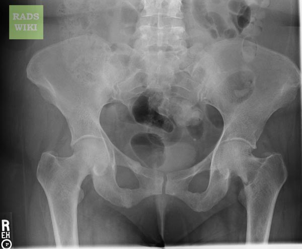
Looser fracture is considered to be a type of insufficiency fracture. On imaging,radiolucent areas occurring at right angles to the cortex and extending across a portion of the bone diameter are seen. It is frequently bilateral. Characteristic sites of involvement are the axillary margins of the scapula, ribs, pubic rami, proximal end of the femur, and ulna.
Causes
- Osteomalacia
- Chronic renal disease
- Fibrous dysplasia
- Hyperthyroidism
- Paget's disease
- Renal osteodystrophy
- X linked hypophosphataemia

