Lateral lemniscus
Template:Infobox Brain Editor-In-Chief: C. Michael Gibson, M.S., M.D. [1]
The lateral lemniscus is a tract of axons in the brainstem that carries information about sound from the cochlear nucleus to various brainstem nuclei and ultimately the contralateral inferior colliculus of the midbrain.
Connections
The brainstem nuclei include:
- the superior olive
- the intermediate nucleus of the lateral lemniscus (INLL)
- the ventral nucleus of the lateral lemniscus (VNLL)
- the dorsal nucleus of the lateral lemniscus (DNLL)
Fibers leaving these brainstem nuclei ascending to the inferior colliculus rejoin the lateral lemniscus. In that sense, this is not a 'lemniscus' in the true sense of the word (second order, decussated sensory axons), as there is third (and out of the lateral superior olive, fourth) order information coming out of some of these brainstem nuclei.
The lateral lemniscus is located where the cochlear nuclei and the pontine reticular formation (PRF) crossover. The PRF descends the reticulospinal tract where it innervates motor neurons and spinal interneurons. It is the main auditory tract in the brainstem which connects the superior olivary complex (SOC) with the inferior colliculus (IC). The Dorsal Cochlear Nucleus (DCN) has input from the LL and output to the contralateral LL via the ipsilateral and contralateral Dorsal Acoustic Stria.
There are three small nuclei on each of the lateral lemnisci; the ventral, dorsal and the intermediate. The two lemnisci communicate via the commissural fibres of Probst.
Function of this structure
This part of the brain also functions in identifying the onset of the sound, the duration of the sound and also with monaural volume.
The function of the lateral lemniscus is not known; however it has good temporal resolution compared to other cells higher than the cochlear nuclei and is sensitive to both timing and amplitude changes in sound. It is also involved in the acoustic startls reflex; the most likely region for this being the VNLL.
The cells of the DNLL respond best to bilateral inputs. Sound in the contralateral ear leads to the strongest responses in the VNLL which deals with some temporary processing. The VNLL may also be essential to the IC’s decoding of amplitude modulated sounds.
DNLL cells have onset and complexity tuned sustained responses. VNLL cells have little spontaneous activinty, broad and moderately complex tuning curves; they have both phasic and tonic responses and are involved in temporal processing.
INLL also has little spontaneous activity and broad tuning curves. The temporal responses are significantly different to cells of the VNLL.
Inputs and outputs to nuclei
The table below shows that each of the nuclei have a complicated arrangement of ipsilateral and contralateral afferent inputs and outputs.
| Nucleus | Input | Output | ||
| Contralateral | Ipsilateral | Contralateral | Ipsilateral | |
| VNLL | Anterior and posterior ventral cochlear nuclei | Medial nucleus of the trapezoid body | Inferior
Colliculus DNLL |
|
| INLL | Anterior and posterior Ventral Cochlear Nucleus | Medial nucleus of the trapezoid body | Medial
Geniculate body Inferior Colliculus |
|
| DNLL | Anterior
Ventral Cochlear nucleus (and Bilateral) |
Medial
superior Olivary Nucleus Lateral Superior Olivary Nucleus (and Bilateral) |
DNLL Inferior Colliculus Mid brain reticular formation Superior Olivary Complex |
Inferior
Colliculus Medial Geniculate Body Mid brain reticular formation Superior Olivary Complex |
Additional images
-
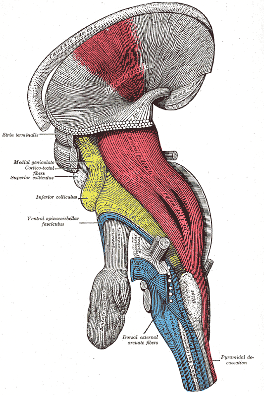
 Dissection of brain-stem. Lateral view.
 -
 Deep dissection of brain-stem. Lateral view.
 -
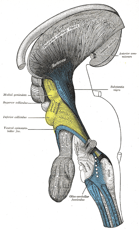 Deep dissection of brain-stem. Lateral view.
 -
 Deep dissection of brain-stem. Ventral view.
 -
 Dissection of brain-stem. Dorsal view.
 -
 Coronal section through mid-brain.
 -
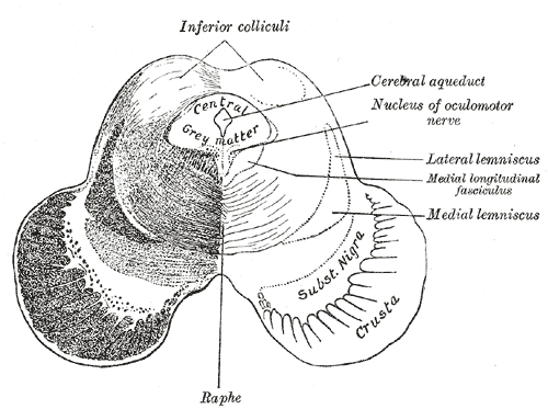 Transverse section of mid-brain at level of inferior colliculi.
 -
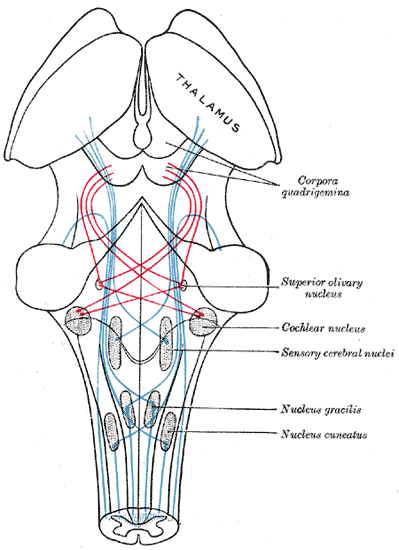 Scheme showing the course of the fibers of the lemniscus; medial lemniscus in blue, lateral in red.


