Kidney stone physical examination
Jump to navigation
Jump to search
|
Kidney stone Microchapters |
|
Diagnosis |
|---|
|
Treatment |
|
Case Studies |
|
Kidney stone physical examination On the Web |
|
American Roentgen Ray Society Images of Kidney stone physical examination |
|
Risk calculators and risk factors for Kidney stone physical examination |
Editor-In-Chief: C. Michael Gibson, M.S., M.D. [1]; Associate Editor(s)-in-Chief: Amandeep Singh M.D.[2]
Overview
Patients with nephrolithiasis usually appear in pain and trying to achieve a comfortable position. Common physical examination findings of nephrolithiasis include costovertebral angle tenderness, may accompany fever with increased heart rate and respiratory rate. They can have hypoactive bowel sounds due to ileus caused by severe pain. They can have hematuria in microscopic exmaination.
Physical Examination
- Physical examination of patients with nephrolithiasis is usually remarkable for:[1]
Appearance of the Patient
- Patients with nephrolithiasis usually appear in pain.
- Patients tend to move constantly in order to achieve a comfortable position.
Vital Signs
- High-grade / low-grade fever.
- Tachycardia with regular pulse can be seen secondary to pain.
- Tachypnea can be seen.
Skin
- Skin examination of patients with nephrolithiasis is usually normal.
HEENT
- HEENT examination of patients with nephrolithiasis is usually normal.
Neck
- Neck examination of patients with nephrolithiasis is usually normal.
Lungs
- Pulmonary examination of patients with nephrolithiasis is usually normal.
Heart
 |
- Cardiovascular examination of patients with nephrolithiasis is usually normal.
Abdomen
- Hypoactive bowel sounds as seen in ileus due to severe pain
Back
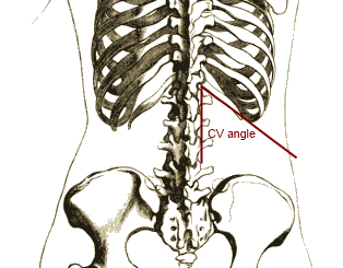
- Costovertebral angle tenderness(Murphy's punch sign, Pasternacki's sign, or Goldflam's sign) bilaterally/unilaterally depending upon sides involved.
Genitourinary
Neuromuscular
- Neuromuscular examination of patients with nephrolithiasis is usually normal.
Extremities
- Extremities examination of patients with nephrolithiasis is usually normal.
References
- ↑ Worcester EM, Coe FL (June 2008). "Nephrolithiasis". Prim. Care. 35 (2): 369–91, vii. doi:10.1016/j.pop.2008.01.005. PMC 2518455. PMID 18486720.
- ↑ https://commons.wikimedia.org/w/index.php?curid=19887470