Gut-associated lymphoid tissue
Editor-In-Chief: C. Michael Gibson, M.S., M.D. [1]
Overview
The digestive tract's immune system is often referred to as gut-associated lymphoid tissue (GALT) and works to protect the body from invasion. GALT is an example of mucosa-associated lymphoid tissue.
Function
About 70% of the body's immune system is found in the digestive tract. The GALT is made up of several types of lymphoid tissue that produce and store immune cells that carry out attacks and defend against pathogens.
New research indicates that GALT may continue to be a major site of HIV activity, even if drug treatment has reduced HIV count in the peripheral blood.[citation needed]
Components
Lymphoid tissue in the gut is comprised of the following :
- Tonsils (Waldeyer's ring)
- Adenoids (Pharyngeal tonsils)
- Peyer's patches
- Lymphoid aggregates in the appendix and large intestine
- Lymphoid tissue accumulating with age in the stomach
- Small lymphoid aggregates in the oesophagus
- Diffusely distributed lymphoid cells and plasma cells in the lamina propria of the gut
Additional images
-
 Lymphatics of colon.
 -
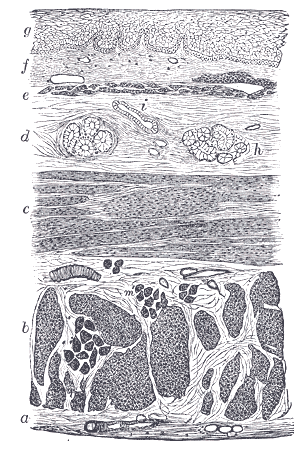 Section of the human esophagus.
 -
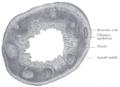 Transverse section of human vermiform process. X 20.
 -
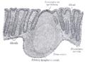 Section of mucous membrane of human rectum. X 60.


External links
- Histology image: 12502loa – Histology Learning System at Boston University - "Digestive System: Alimentary Canal: colon, taenia coli"
- Histology image: 11102loa – Histology Learning System at Boston University - "Digestive System: Alimentary Canal: esophageal/stomach junction"
de:Darmassoziiertes Immunsystem Template:Digestive tract Template:Lymphatic system Template:WikiDoc Sources