Great saphenous vein
Editor-In-Chief: C. Michael Gibson, M.S., M.D. [1]
The great saphenous vein, also greater saphenous vein, is the large (subcutaneous) superficial vein of the leg and thigh.
Path
It originates from where the dorsal vein of the first digit (the large toe) merges with the dorsal venous arch of the foot.
After passing anterior to the medial malleolus (where it often can be visualized and palpated), it runs up the medial side of the leg.
At the knee, it runs over the posterior border of the medial epicondyle of the femur bone.
The great saphenous vein then courses laterally to lie on the anterior surface of the thigh before entering an opening in the fascia lata called the saphenous opening.
It joins with the femoral vein in the region of the femoral triangle.
Tributaries
At the ankle it receives branches from the sole of the foot through the medial marginal vein; in the lower leg it anastomoses freely with the small saphenous vein, communicates with the anterior and posterior tibial veins and receives many cutaneous veins; in the thigh it communicates with the femoral vein and receives numerous tributaries; those from the medial and posterior parts of the thigh frequently unite to form a large accessory saphenous vein which joins the main vein at a variable level.
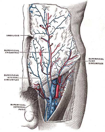
Near the fossa ovalis it is joined by the superficial epigastric, superficial iliac circumflex, and superficial external pudendal veins.
A vein, named the thoracoepigastric, runs along the lateral aspect of the trunk between the superficial epigastric vein below and the lateral thoracic vein above and establishes an important communication between the femoral and axillary veins.
Use in cardiovascular procedures
The vein is often removed by cardiac surgeons and used for autotransplantation in coronary artery bypass operations, when arterial grafts are not available or many grafts are required, such as in a triple bypass or quadruple bypass.
The great saphenous vein is the conduit of choice for vascular surgeons,[1][2] when available, for doing peripheral arterial bypass operations because it has superior long-term patency compared to synthetic grafts (PTFE, PETE (Dacron®)), human umbilical vein grafts or biosynthetic grafts [Omniflow®]. Often, it is used in situ (in place), after tying off smaller tributaries and stripping the valves with a device called LeMaitre's valvulotome.
The saphenous nerve is a branch of the femoral nerve that runs with the great saphenous vein and is often damaged in surgeries that make use of the similarly named vein.
Pathology of the saphenous vein
Pathology of the great saphenous vein is relatively common, but in isolation typically not life threatening.[3]
- Varicose veins: The great saphenous vein, like other superficial veins, can develop varices, which are generally considered to be unsightly. Various treatment options exist for treating varicose veins. Varicose veins are not life threatening.
- Thrombophlebitis: The great saphenous vein can thrombose and become infected. Thrombophlebitis of the great saphenous vein is not life threatening in isolation; however, it may be associated with deep vein thrombosis which can be and thus requires further investigation.[3]
See also
Additional images
-
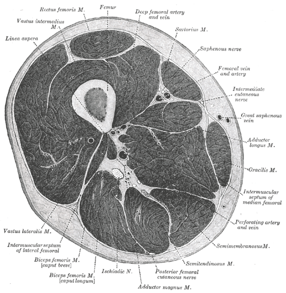 Cross-section through the middle of the thigh.
 -
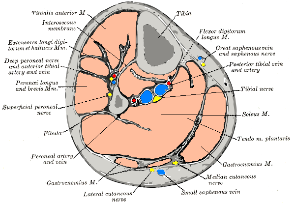 Cross-section through middle of leg.
 -
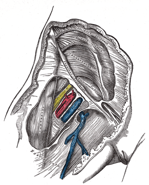 Femoral sheath laid open to show its three compartments.
 -
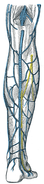 Small saphenous vein and its tributaries.
 -
 The femoral vein and its tributaries.


References
- ↑ Muhs BE, Gagne P, Sheehan P. Peripheral arterial disease: clinical assessment and indications for revascularization in the patient with diabetes. Curr Diab Rep. 2005 Feb;5(1):24-9. PMID 15663913.
- ↑ Mamode N, Scott RN. Graft type for femoro-popliteal bypass surgery. Cochrane Database Syst Rev. 2000;(2):CD001487. PMID 10796649.
- ↑ 3.0 3.1 Feied C, Handler JA. Thrombophlebitis, Superficial. eMedicine.com. Available at: http://www.emedicine.com/emerg/topic582.htm. Accessed on: December 18, 2006.
External links
- Template:GraySubject - "The arteries of the lower extremity" - Gray's Anatomy.
- Template:GraySubject - "The veins of the lower extremity, abdomen, and pelvis" - Gray's Anatomy.
- Great saphenous vein - Stedman's medical dictionary.
- Template:SUNYAnatomyLabs
- Template:SUNYAnatomyLabs
- Template:MedicalMnemonics