Flexor hallucis longus muscle
Overview
The Flexor hallucis longus muscle (FHL) is a muscle of the leg. It is one of the deep muscles of the posterior compartment of the leg. the other deep muscles of the leg are flexor digitorum longus and tibialis posterior. FHL is the largest and most powerful of these deep muscles.
The Flexor hallucis longus is situated on the fibular side of the leg. It arises from the inferior two-thirds of the posterior surface of the body of the fibula, with the exception of 2.5 cm. at its lowest part; from the lower part of the interosseous membrane; from an intermuscular septum between it and the Peronæi, laterally, and from the fascia covering the Tibialis posterior, medially.
The fibers pass obliquely downward and backward, and end in a tendon which occupies nearly the whole length of the posterior surface of the muscle.
This tendon lies in a groove which crosses the posterior surface of the lower end of the tibia, the posterior surface of the talus, and the under surface of the sustentaculum tali of the calcaneus; in the sole of the foot it runs forward between the two heads of the Flexor hallucis brevis, and is inserted into the base of the last phalanx of the great toe. The grooves on the talus and calcaneus, which contain the tendon of the muscle, are converted by tendinous fibers into distinct canals, lined by a mucous sheath.
As the tendon passes forward in the sole of the foot, it is situated above, and crosses from the lateral to the medial side of the tendon of the Flexor digitorum longus, to which it is connected by a fibrous slip.
Variations
Usually a slip runs to the Flexor digitorum and frequently an additional slip runs from the Flexor digitorum to the Flexor hallucis. Peroneocalcaneus internus, rare, origin below or outside the Flexor hallucis from the back of the fibula, passes over the sustentaculum tali with the Flexor hallucis and is inserted into the calcaneum.
Additional images
-
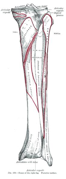 Bones of the right leg. Posterior surface.
 -
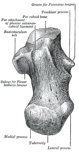 Left calcaneus, inferior surface.
 -
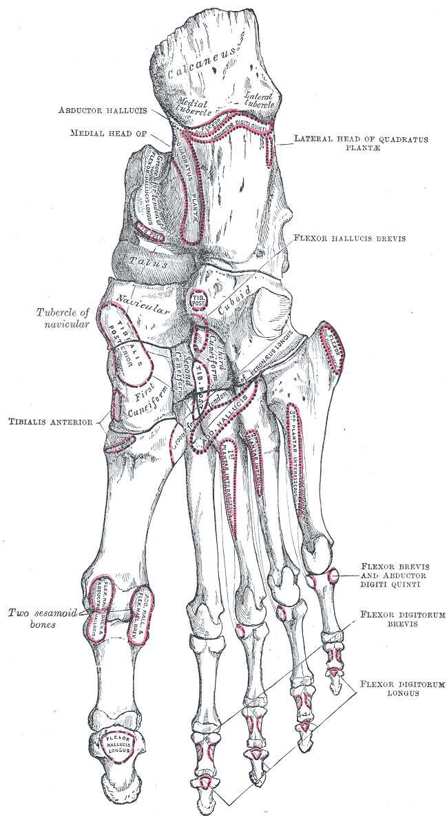 Bones of the right foot. Plantar surface.
 -
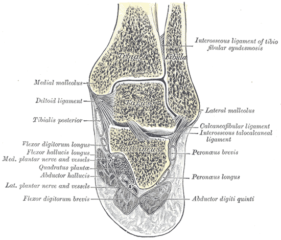 Coronal section through right talocrural and talocalcaneal joints.
 -
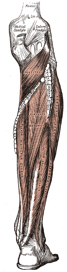 Muscles of the back of the leg. Deep layer.
 -
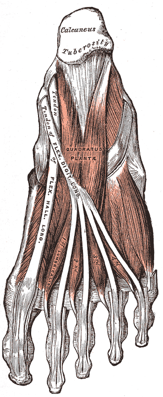 Muscles of the sole of the foot. Second layer.
 -
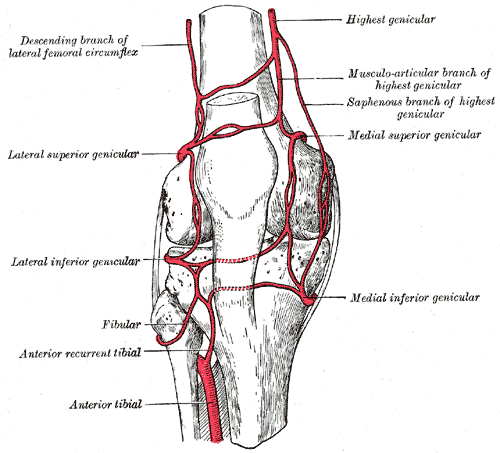
 The popliteal, posterior tibial, and peroneal arteries.
 -
 Circumpatellar anastomosis.
 -
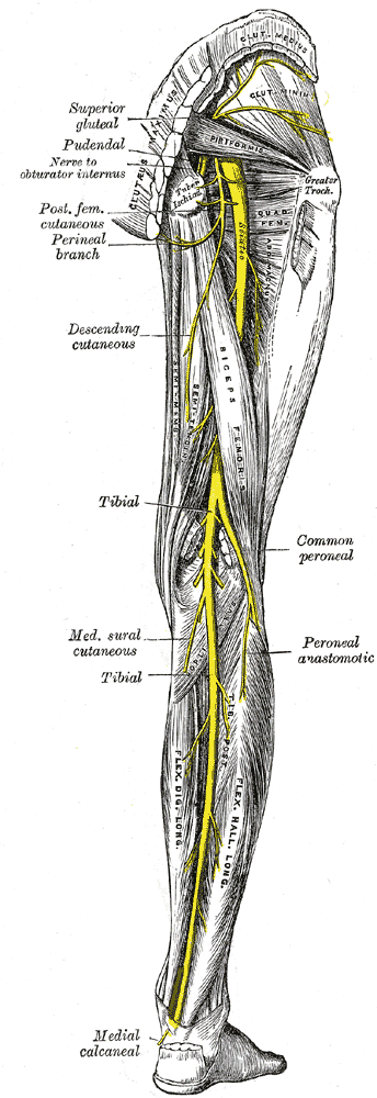 Nerves of the right lower extremity Posterior view.
 -
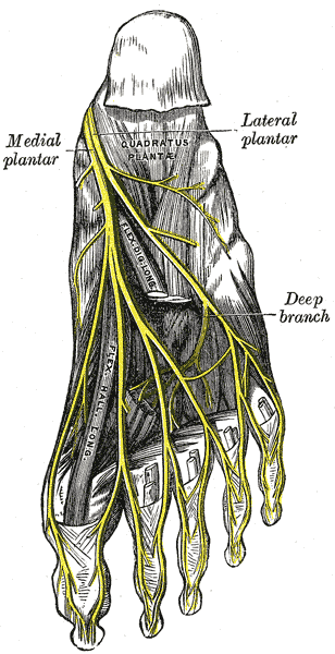 The plantar nerves.


External links
Template:Gray's Template:Muscles of lower limb
de:Musculus flexor hallucis longus sv:Flexor hallucis longus Template:WH Template:WikiDoc Sources