Enterobiasis laboratory findings
|
Enterobiasis Microchapters |
|
Diagnosis |
|---|
|
Treatment |
|
Case Studies |
|
Enterobiasis laboratory findings On the Web |
|
American Roentgen Ray Society Images of Enterobiasis laboratory findings |
|
Risk calculators and risk factors for Enterobiasis laboratory findings |
Editor-In-Chief: C. Michael Gibson, M.S., M.D. [1] Associate Editor(s)-in-Chief: Furqan M M. M.B.B.S[2]
Overview
Diagnosis of enterobiasis is often made clinically by observing the female worm(s) in the peri-anal region, but can also be made using the "scotch-tape" test, in which the sticky side of a strip of cellophane tape is pressed against the peri-anal skin, then examined under a microscope for pinworm eggs.
Laboratory Findings
Scotch Tape Test
It is also called Hall or National Institute of Health swab. Sticky side of a strip of cellophane tape is pressed against the peri-anal skin, then examined under a microscope for pinworm eggs. The test is repeated for five consecutive mornings to increase the sensitivity to 99%. It is done prior to washing or defecation. The diagnostic characteristics of egg are: size 50-54 µm by 20-27 µm; typical elongated shape, with one convex side and one flattened side and colorless shell.[1][2]
Stool analysis
Stool analysis for ova and parasites is of low diagnostic yield. The actual worms may be seen in the host's feces; however the eggs are invisible to the naked eye.[1]
Histology
On histologic cross-section alae or wings (running the length of the worm) are identifying features of the pinworm (see micrograph).[3]
Images
-
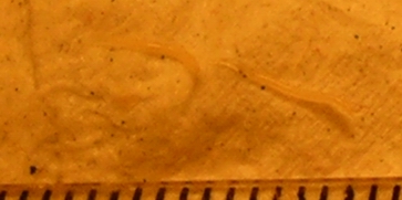 Two pinworms, captured on emergence from the anus. Markings are 1 mm apart.
 -
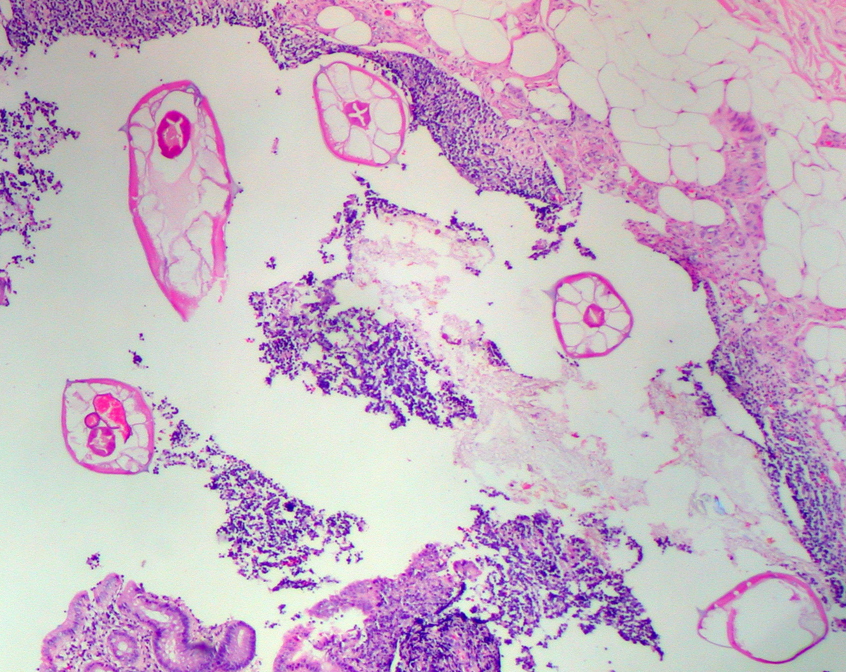 Pinworms are sometimes diagnosed incidentally by pathology. Micrograph of pinworms in the appendix. H&E stain.
 -
-
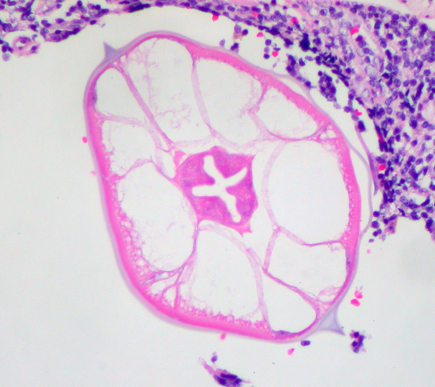
 High magnification micrograph of a pinworm in cross-section in the appendix. H&E stain.
 -
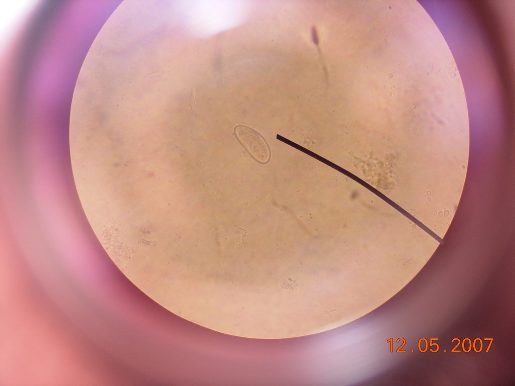 Enterobius vermicularis egg under a light microscope.
 -
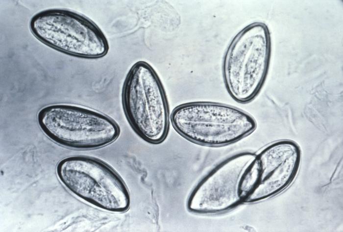 Pinworm eggs are easily seen under a microscope.
 -
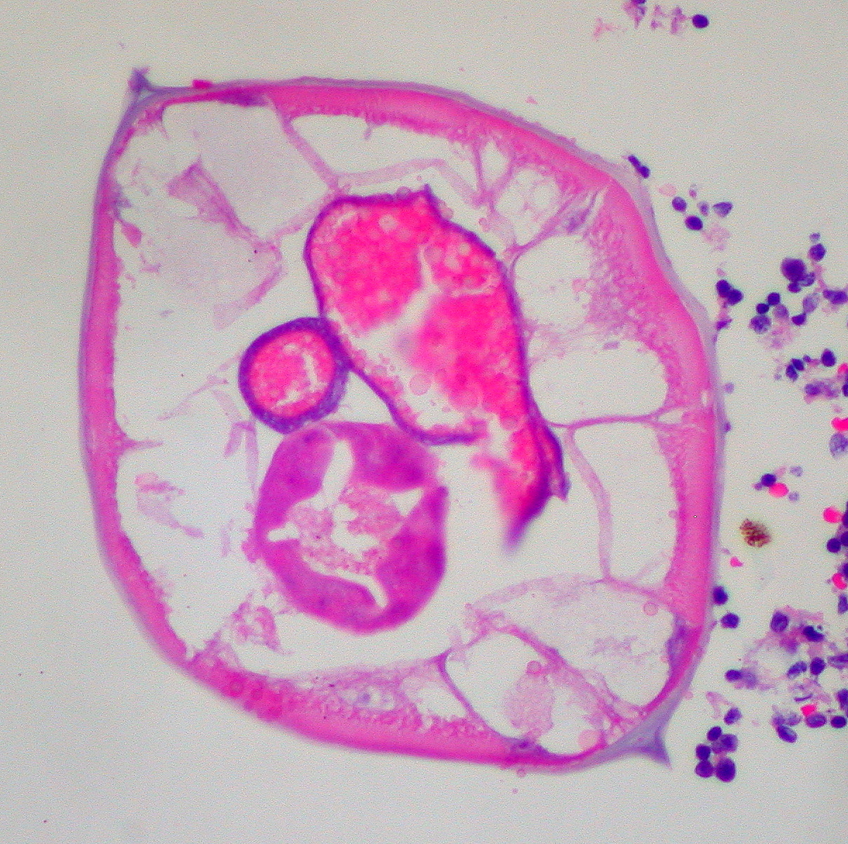
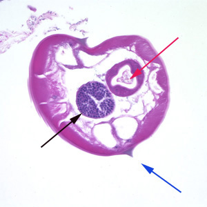 Pinworms are sometimes diagnosed incidentally by pathology. Micrograph of male pinworm in cross-section. Alae (blue arrow), intestine (red arrow) and testis (black arrow). H&E stain.
 -
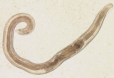 A pinworm (Enterobius vermicularis).

.jpg)
.jpg)
.jpg)


Videos Showing Pinworm Egg Under Microscope
{{#ev:youtube|YNrkWStDdmo}}
References
- ↑ 1.0 1.1 Caldwell JP (1982). "Pinworms (enterobius vermicularis)". Can Fam Physician. 28: 306–9. PMC 2306321. PMID 21286054.
- ↑ Cook GC (1994). "Enterobius vermicularis infection". Gut. 35 (9): 1159–62. PMC 1375686. PMID 7959218.
- ↑ Diagnostic Findings Enterobiasis. Centers for Disease Control and Prevention. URL:http://www.dpd.cdc.gov/dpdx/HTML/Enterobiasis.htm. Accessed on: August 6, 2008.