Diabetic retinopathy physical examination
|
Diabetic retinopathy Microchapters |
|
Diagnosis |
|---|
|
Treatment |
|
Case Studies |
|
Diabetic retinopathy physical examination On the Web |
|
American Roentgen Ray Society Images of Diabetic retinopathy physical examination |
|
Risk calculators and risk factors for Diabetic retinopathy physical examination |
Editor-In-Chief: C. Michael Gibson, M.S., M.D. [1]
Overview
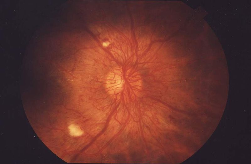
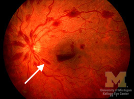
On fundoscopic exam, diabetic retinopathy is characterized by cotton wool spots, flame hemorrhages, dot-blot hemorrhages and boat hemorrhages.
Physical Examination
- Visual acuity test: This eye chart test measures how well you see at various distances.
- Dilated eye exam: Drops are placed in your eyes to widen, or dilate, the pupils. Your eye care professional uses a special magnifying lens to examine your retina and optic nerve for signs of damage and other eye problems. After the exam, your close-up vision may remain blurred for several hours.
Ophthalmoscopic findings
Cotton Wool Spots

Cotton wool spots are an abnormal finding on fundoscopic exam of the retina. They appear as puffy white patches on the retina. They are caused by damage to nerve fibers. The nerve fibers are damaged by swelling in the surface layer of the retina. The cause of this swelling is due to the reduced axonal transport (and hence backlog of intracellular products) within the nerves because of the ischaemia.
Flame Hemorrhages

Flame hemorrhages are flame shaped hemorrhages located in the superficial nerve fiber layer of the retina that appear dark dark red on fundoscopic examination. Flame hemorrhages are caused by leakage from arterioles due to ischemic damage or from veins that are ischemic or in under high pressure.
Dot Hemorrhages

Dot hemorrhages are dark red round spots of hemorrhage seen on fundoscopic exam. They are frequently observed in patients with diabetic retinopathy. Dot hemorrhages are due to either capillary or venular leak. The site of hemorrhage is deep within the retina.
Boat Hemorrhages

Boat hemorrhages are rectangular dark red spots of hemorrhage seen on fundoscopic exam. They are frequently observed in patients with diabetic retinopathy. Boat hemorrhages are due to either capillary or venular leak. The site of hemorrhage is at the interface between the retina and the vitreous humor. The contents that leak out are under such high-pressure that they break through the internal liminiting membrane of the retina.