Cyclops lesion
|
WikiDoc Resources for Cyclops lesion |
|
Articles |
|---|
|
Most recent articles on Cyclops lesion Most cited articles on Cyclops lesion |
|
Media |
|
Powerpoint slides on Cyclops lesion |
|
Evidence Based Medicine |
|
Clinical Trials |
|
Ongoing Trials on Cyclops lesion at Clinical Trials.gov Trial results on Cyclops lesion Clinical Trials on Cyclops lesion at Google
|
|
Guidelines / Policies / Govt |
|
US National Guidelines Clearinghouse on Cyclops lesion NICE Guidance on Cyclops lesion
|
|
Books |
|
News |
|
Commentary |
|
Definitions |
|
Patient Resources / Community |
|
Patient resources on Cyclops lesion Discussion groups on Cyclops lesion Patient Handouts on Cyclops lesion Directions to Hospitals Treating Cyclops lesion Risk calculators and risk factors for Cyclops lesion
|
|
Healthcare Provider Resources |
|
Causes & Risk Factors for Cyclops lesion |
|
Continuing Medical Education (CME) |
|
International |
|
|
|
Business |
|
Experimental / Informatics |
Editor-In-Chief: C. Michael Gibson, M.S., M.D. [1]
Overview
The cyclops lesion occurs with an estimated frequency of 1%–9.8% of patients following anterior cruciate ligament reconstruction. Although the precise cause is unknown, it is believed that uplifting of fibrocartilaginous tissue during drilling of the tibia for anterior cruciate ligament reconstruction serves as a nidus for fibrous tissue deposition. Pathologically, the lesion consists of central granulation tissue surrounded by dense fibrous tissue. The lesion was so named because of its bulbous appearance and characteristic focal areas of reddish-blue discoloration (from venous channels) that resemble an eye at arthroscopy. The lesion may result in loss of full extension and surgical débridement of the lesion results in restoration of full knee extension.
Diagnosis
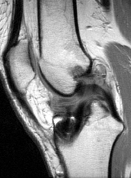
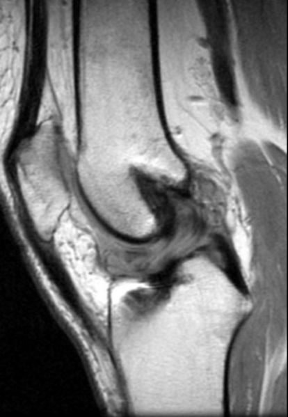
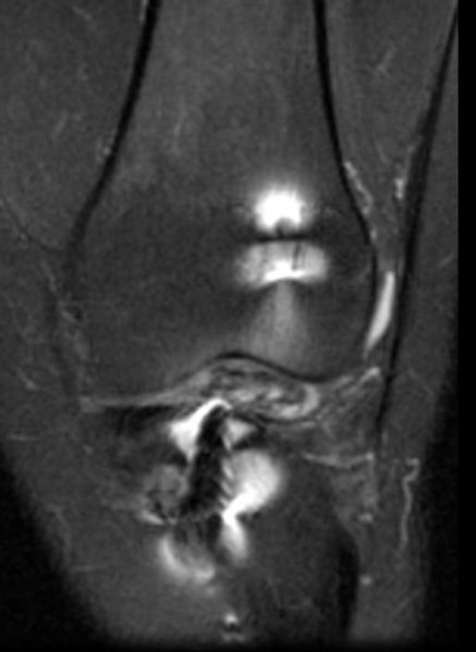
- At MR imaging, a soft-tissue mass is seen anteriorly or anterolaterally in the intercondylar notch near the tibial insertion of the reconstructed anterior cruciate ligament.
- Because of its fibrous content, a cyclops lesion typically has intermediate to low signal intensity with all pulse sequences.
Disruption of ACL graft with cyclops lesion


