Anterior corticospinal tract
| Anterior corticospinal tract | |
|---|---|
| Anterior corticospinal tract is 1b, in red at bottom center. | |
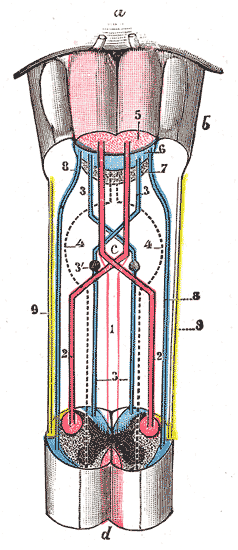
| Decussation of pyramids.
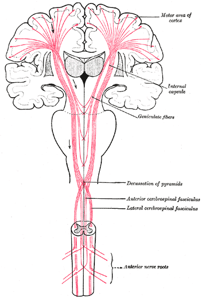
 Scheme showing passage of various fasciculi from medulla spinalis to medulla oblongata. a. Pons. b. Medulla oblongata. c. Decussation of the pyramids. d. Section of cervical part of medulla spinalis. 1. Anterior cerebrospinal fasciculus (in red). 2. Lateral cerebrospinal fasciculus (in red). 3. Sensory tract (fasciculi gracilis et cuneatus) (in blue). 3’. Gracile and cuneate nuclei. 4. Antero-lateral proper fasciculus (in dotted line). 5. Pyramid. 6. Lemniscus. 7. Medial longitudinal fasciculus. 8. Ventral spinocerebellar fasciculus (in blue). 9. Dorsal spinocerebellar fasciculus (in yellow). | |
| Latin | tractus corticospinalis anterior, fasciculus cerebrospinalis anterior |
| Gray's | subject #185 759 |
| Dorlands/Elsevier | t_15/12816937 |
Overview
The anterior corticospinal tract (also called the ventral corticospinal tract, medial corticospinal tract, direct pyramidal tract, or anterior cerebrospinal fasciculus) is a small bundle of descending fibers that connect the cerebral cortex to the spinal cord. It is usually small, varying inversely in size with the lateral corticospinal tract, which is the main part of the corticospinal tract.
It lies close to the anterior median fissure, and is present only in the upper part of the medulla spinalis; gradually diminishing in size as it descends, it ends about the middle of the thoracic region.
It consists of descending fibers which arise from cells in the motor area of the cerebral hemisphere of the same side, and which, as they run downward in the medulla spinalis, cross in succession through the anterior white commissure to the opposite side, where they end, either directly or indirectly, by arborizing around the motor cells in the anterior column.
A few of its fibers are said to pass to the lateral column of the same side and to the gray matter at the base of the posterior column.
They conduct voluntary motor impulses from the precentral gyrus to the motor centers of the cord.
Additional images
External links
- NeuroNames hier-799
- 745209857 at GPnotebook
- Anterior+corticospinal+tract at eMedicine Dictionary
- Overview at thebrain.mcgill.ca
This article was originally based on an entry from a public domain edition of Gray's Anatomy. As such, some of the information contained herein may be outdated. Please edit the article if this is the case, and feel free to remove this notice when it is no longer relevant.
Brain: rhombencephalon (hindbrain) | |
|---|---|
| Myelencephalon/medulla | anterior/ventral: Arcuate nucleus of medulla • Pyramid (Decussation) • Olivary body • Inferior olivary nucleus • Anterior median fissure • Ventral respiratory group posterior/dorsal: VII,IX,X: Solitary/tract • XII, X: Dorsal • IX,X,XI: Ambiguus • IX: Inferior salivatory nucleus • Gracile nucleus/Cuneate nucleus/Accessory cuneate nucleus • Area postrema • Posterior median sulcus • Dorsal respiratory group raphe/reticular: Sensory decussation • Reticular formation (Gigantocellular nucleus, Parvocellular reticular nucleus, Ventral reticular nucleus, Lateral reticular nucleus, Paramedian reticular nucleus) • Raphe nuclei (Obscurus, Magnus, Pallidus) tracts: Corticospinal tract (Lateral, Anterior) • Inferior cerebellar peduncle • Olivocerebellar tract • Spinocerebellar (Dorsal, Ventral) • Spinothalamic tract • PCML (Posterior external arcuate fibers, Internal arcuate fibers, Medial lemniscus) • Extrapyramidal (Rubrospinal tract, Vestibulospinal tract, Tectospinal tract) |
| Metencephalon/pons | anterior/ventral: Superior olivary nucleus • Basis pontis (Pontine nuclei, Middle cerebellar peduncles) posterior/dorsal: Pontine tegmentum (Trapezoid body, Superior medullary velum, Locus ceruleus, MLF, Vestibulocerebellar tract, V Principal Spinal & Motor, VI, VII, VII: Superior salivary nucleus) • VIII-c (Dorsal, Anterior)/VIII-v (Lateral, Superior, Medial, Inferior) raphe/reticular: Reticular formation (Caudal pontine reticular nucleus, Oral pontine reticular nucleus, Tegmental pontine reticular nucleus, Paramedian pontine reticular formation) • Median raphe nucleus |
| Metencephalon/cerebellum | Vermis • Flocculus • Arbor vitae • Cerebellar tonsil • Inferior medullary velum Molecular layer (Stellate cell, Basket cell, Parallel fiber) • Purkinje cell layer (Purkinje cell) • Granule cell layer (Golgi cell) • Mossy fibers • Climbing fiber |
| Fourth ventricle | apertures (Median, Lateral) • Rhomboid fossa (Vagal trigone, Hypoglossal trigone, Obex, Sulcus limitans, Facial colliculus, Medial eminence) • Lateral recess |
Table of Contents In Alphabetical Order | By Individual Diseases | Signs and Symptoms | Physical Examination | Lab Tests | Drugs
Editor Tools Become an Editor | Editors Help Menu | Create a Page | Edit a Page | Upload a Picture or File | Printable version | Permanent link | Maintain Pages | What Pages Link HereThere is no pharmaceutical or device industry support for this site and we need your viewer supported Donations | Editorial Board | Governance | Licensing | Disclaimers | Avoid Plagiarism | Policies

