Alopecia mucinosa
|
WikiDoc Resources for Alopecia mucinosa |
|
Articles |
|---|
|
Most recent articles on Alopecia mucinosa Most cited articles on Alopecia mucinosa |
|
Media |
|
Powerpoint slides on Alopecia mucinosa |
|
Evidence Based Medicine |
|
Cochrane Collaboration on Alopecia mucinosa |
|
Clinical Trials |
|
Ongoing Trials on Alopecia mucinosa at Clinical Trials.gov Trial results on Alopecia mucinosa Clinical Trials on Alopecia mucinosa at Google
|
|
Guidelines / Policies / Govt |
|
US National Guidelines Clearinghouse on Alopecia mucinosa NICE Guidance on Alopecia mucinosa
|
|
Books |
|
News |
|
Commentary |
|
Definitions |
|
Patient Resources / Community |
|
Patient resources on Alopecia mucinosa Discussion groups on Alopecia mucinosa Patient Handouts on Alopecia mucinosa Directions to Hospitals Treating Alopecia mucinosa Risk calculators and risk factors for Alopecia mucinosa
|
|
Healthcare Provider Resources |
|
Causes & Risk Factors for Alopecia mucinosa |
|
Continuing Medical Education (CME) |
|
International |
|
|
|
Business |
|
Experimental / Informatics |
Editor-In-Chief: C. Michael Gibson, M.S., M.D. [1]; Mugilan Poongkunran M.B.B.S [2], Jesus Rosario Hernandez, M.D. [3].
Synonyms and keywords: Follicular mucinosis
Overview
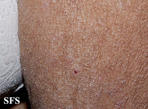
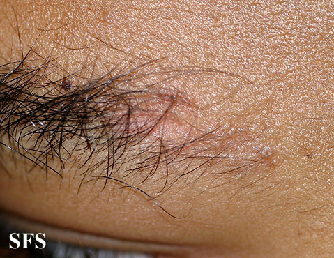
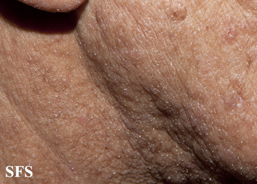
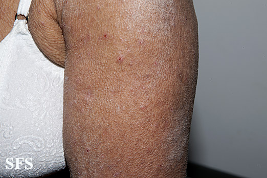
Alopecia mucinosa is an epithelial reaction pattern that is characterized by the accumulation of mucinous material in the epithelial hair follicle sheath and the sebaceous glands. This generally presents, but not exclusively, as erythematous plaques or flat patches without hair primarily on the scalp and face.[1][2] This can also present on the body as a follicular mucinosis and may represent a systemic disease.[3][4]
Historical Perspective
Pinkus in 1957 first described the term "alopecia mucinosa" for 6 cases of localized alopecia that were histopathologically characterized by mucin deposition within the hair follicles. Follicular mucinosis was later suggested as a better term since the clinical features are variable and alopecia is not always observed.[5]
Classification
Follicular mucinosis is classified into two types:
- The primary type occurs when there is no underlying associated skin disease.
- The secondary type is associated with a number of inflammatory disorders and malignant conditions, including mycosis fungoides and other lymphomas.
Pathophysiology
It has been suggested that the keratinocytes that form the affected follicles produce intracellular mucin and they eventually degenerate, which is induced by the T lymphocytes of the infiltrate.[6]
Causes
The etiology of alopecia mucinosa is unknown. The self-healing course and the pathological picture suggest a virus infection.
Differentiating Alopecia Mucinosa from other Conditions
Alopecia mucinosa need to be differentiated from other follicular papular eruptions such as
- Keratosis pilaris
- Lichen nitidus
- Lichen spinulosus
- Lichen scrofulosorum
Other conditions associated with alopecia need to be differentiated to make a clear diagnosis.
Diagnosis
History and Symptoms
Patients are usually asymptomatic.
Physical Findings
- The most prominent lesion is follicular papules. These may be edematous or not and there may or may not be associated erythema.
- Follicular papules may contain hair often surrounded by sheath of hyperkertosis. These remaining hairs are often distorted and clubbed.
- Also present with poorly demarcated alopecia on the eyebrows, left parietal scalp and pubic area.
Physical examination
Gallery
Skin
Head
Extremities
Trunk
Laboratory Findings
Histopathological examination of a biopsy from lesion would reveal the following:
- Reticular epithelial degeneration and areas of cavitation within the pilosebaceous units and
- Lymphocytes, histiocytes and many eosinophils infiltrated around and into the hair follicles and the sebaceous glands.
Treatment
- Indomethacin, interferon alpha and interferon gamma are the latest used for the treatment.[7][8]
- Steroids, minocycline, and dapsone are turing out to be ineffective in the management of alopecia mucinosa.
- The lesions may also respond to small doses of irradiation.
References
- ↑ Freedberg, et al. (2003). Fitzpatrick's Dermatology in General Medicine. (6th ed.). McGraw-Hill. ISBN 0-07-138076-0.
- ↑ James, William D.; Berger, Timothy G.; et al. (2006). Andrews' Diseases of the Skin: clinical Dermatology. Saunders Elsevier. ISBN 0-7216-2921-0.
- ↑ Rapini, Ronald P.; Bolognia, Jean L.; Jorizzo, Joseph L. (2007). Dermatology: 2-Volume Set. St. Louis: Mosby. ISBN 1-4160-2999-0.
- ↑ Folliculitis, follicular mucinosis, and papular mucinosis as a presentation of chronic myelomonocytic leukemia. Rashid R, Hymes S. Dermatol Online J. 2009 May 15;15(5):16.
- ↑ JABLONSKA S, CHORZELSKI T, LANCUCKI J (1959). "[Mucinosis follicularis]". Hautarzt. 10: 27–33. PMID 14406240.
- ↑ Reed RJ (1981). "The T-lymphocyte, the mucinous epithelial interstitium, and immunostimulation". Am J Dermatopathol. 3 (2): 207–14. PMID 6973937.
- ↑ Kodama H, Umemura S, Nohara N (1988). "Follicular mucinosis: response to indomethacin". J Dermatol. 15 (1): 72–5. PMID 2969013.
- ↑ Meissner K, Weyer U, Kowalzick L, Altenhoff J (1991). "Successful treatment of primary progressive follicular mucinosis with interferons". J Am Acad Dermatol. 24 (5 Pt 2): 848–50. PMID 1828814.


