Whipworm infection: Difference between revisions
No edit summary |
No edit summary |
||
| Line 24: | Line 24: | ||
The unembryonated eggs are passed with the stool '''1.'''. In the soil, the eggs develop into a 2-cell stage '''2.''', an advanced cleavage stage '''3.''', and then they embryonate '''4.'''; eggs become infective in 15 to 30 days. After ingestion (soil-contaminated hands or food), the eggs hatch in the small intestine, and release larvae '''5.''' that mature and establish themselves as adults in the colon '''6.'''. The adult worms (approximately 4 cm in length) live in the cecum and ascending colon. The adult worms are fixed in that location, with the anterior portions threaded into the mucosa. The females begin to oviposit 60 to 70 days after infection. Female worms in the cecum shed between 3,000 and 20,000 eggs per day. The life span of the adults is about 1 year. | The unembryonated eggs are passed with the stool '''1.'''. In the soil, the eggs develop into a 2-cell stage '''2.''', an advanced cleavage stage '''3.''', and then they embryonate '''4.'''; eggs become infective in 15 to 30 days. After ingestion (soil-contaminated hands or food), the eggs hatch in the small intestine, and release larvae '''5.''' that mature and establish themselves as adults in the colon '''6.'''. The adult worms (approximately 4 cm in length) live in the cecum and ascending colon. The adult worms are fixed in that location, with the anterior portions threaded into the mucosa. The females begin to oviposit 60 to 70 days after infection. Female worms in the cecum shed between 3,000 and 20,000 eggs per day. The life span of the adults is about 1 year. | ||
<br clear="left"/> | |||
==Diagnosis== | ==Diagnosis== | ||
'''Microscopy''' | '''Microscopy''' | ||
[[Image:Trichuris eggA.jpg|thumb|left|T.trichiura egg]] | [[Image:Trichuris eggA.jpg|thumb|left|T.trichiura egg]] | ||
<br clear="left"/> | |||
A: Trichuris trichiura egg (wet preparation). The diagnostic characteristics are: | A: Trichuris trichiura egg (wet preparation). The diagnostic characteristics are: | ||
| Line 39: | Line 39: | ||
[[Image:Trichuris eggC.jpg|thumb|left|Trichuris egg]] | [[Image:Trichuris eggC.jpg|thumb|left|Trichuris egg]] | ||
<br clear="left"/> | |||
C: Trichuris trichiura eggs. Figures show side-by-side eggs with regular (white arrows) and larger (black arrows) size eggs. | C: Trichuris trichiura eggs. Figures show side-by-side eggs with regular (white arrows) and larger (black arrows) size eggs. | ||
[[Image:Trichuris eggD.jpg|thumb|left|Trichuris egg]] | [[Image:Trichuris eggD.jpg|thumb|left|Trichuris egg]] | ||
<br clear="left"/> | |||
D: Atypical Trichuris sp. egg. | D: Atypical Trichuris sp. egg. | ||
{{SIB}} | {{SIB}} | ||
Revision as of 22:35, 18 January 2009
Please Take Over This Page and Apply to be Editor-In-Chief for this topic: There can be one or more than one Editor-In-Chief. You may also apply to be an Associate Editor-In-Chief of one of the subtopics below. Please mail us [1] to indicate your interest in serving either as an Editor-In-Chief of the entire topic or as an Associate Editor-In-Chief for a subtopic. Please be sure to attach your CV and or biographical sketch.
Overview
The nematode (roundworm) Trichuris trichiura, also called the human whipworm.
Epidemiology and Demographics
Demographics
The third most common round worm of humans. Worldwide, with infections more frequent in areas with tropical weather and poor sanitation practices, and among children. It is estimated that 800 million people are infected worldwide. Trichuriasis occurs in the southern United States.
Infections with the soil-transmitted intestinal helminths (i.e., Ascaris lumbricoides, Trichuris trichiura, and hookworm), estimated to affect approximately 1 billion persons, are among the most common and widespread human infections.
Risk Factors
Most frequently asymptomatic. Heavy infections, especially in small children, can cause gastrointestinal problems (abdominal pain, diarrhea, rectal prolapse) and possibly growth retardation.
Pathophysiology & Etiology

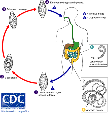
The unembryonated eggs are passed with the stool 1.. In the soil, the eggs develop into a 2-cell stage 2., an advanced cleavage stage 3., and then they embryonate 4.; eggs become infective in 15 to 30 days. After ingestion (soil-contaminated hands or food), the eggs hatch in the small intestine, and release larvae 5. that mature and establish themselves as adults in the colon 6.. The adult worms (approximately 4 cm in length) live in the cecum and ascending colon. The adult worms are fixed in that location, with the anterior portions threaded into the mucosa. The females begin to oviposit 60 to 70 days after infection. Female worms in the cecum shed between 3,000 and 20,000 eggs per day. The life span of the adults is about 1 year.
Diagnosis
Microscopy

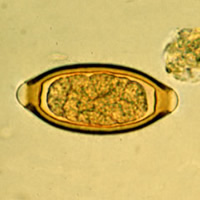
A: Trichuris trichiura egg (wet preparation). The diagnostic characteristics are:
- a typical barrel shape
- two polar plugs, that are unstained
- size: 50 to 54 µm by 22 to 23 µm
The external layer of the shell of the egg is yellow-brown (in contrast to the clear polar plugs). The egg is unembryonated, as eggs are when passed with the stool.

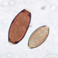
C: Trichuris trichiura eggs. Figures show side-by-side eggs with regular (white arrows) and larger (black arrows) size eggs.

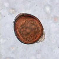
D: Atypical Trichuris sp. egg.