WBR0314: Difference between revisions
No edit summary |
m (refreshing WBR questions) |
||
| (3 intermediate revisions by one other user not shown) | |||
| Line 1: | Line 1: | ||
{{WBRQuestion | {{WBRQuestion | ||
|QuestionAuthor={{Ochuko}} | |QuestionAuthor= {{Ochuko}} (Reviewed by {{YD}}) | ||
|ExamType=USMLE Step 1 | |ExamType=USMLE Step 1 | ||
|MainCategory=Microbiology | |MainCategory=Microbiology | ||
|SubCategory=Pulmonology | |SubCategory=Pulmonology | ||
|MainCategory=Microbiology | |MainCategory=Microbiology | ||
|SubCategory=Pulmonology | |SubCategory=Pulmonology | ||
|MainCategory=Microbiology | |MainCategory=Microbiology | ||
|SubCategory=Pulmonology | |SubCategory=Pulmonology | ||
|MainCategory=Microbiology | |MainCategory=Microbiology | ||
|MainCategory=Microbiology | |MainCategory=Microbiology | ||
|MainCategory=Microbiology | |MainCategory=Microbiology | ||
|SubCategory=Pulmonology | |SubCategory=Pulmonology | ||
|MainCategory=Microbiology | |MainCategory=Microbiology | ||
|SubCategory=Pulmonology | |SubCategory=Pulmonology | ||
|MainCategory=Microbiology | |MainCategory=Microbiology | ||
|SubCategory=Pulmonology | |SubCategory=Pulmonology | ||
|MainCategory=Microbiology | |MainCategory=Microbiology | ||
|SubCategory=Pulmonology | |SubCategory=Pulmonology | ||
|MainCategory=Microbiology | |MainCategory=Microbiology | ||
|MainCategory=Microbiology | |MainCategory=Microbiology | ||
|SubCategory=Pulmonology | |SubCategory=Pulmonology | ||
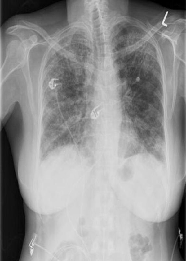
|Prompt=A 22-year old | |Prompt=A 22-year-old female military recruit presents to the emergency department (ED) in early November with complaints of malaise, persistent dry cough, and intermittent low-grade fever for the past 3 weeks. She states that although she is generally able to tolerate her symptoms, her cough has worsened over time. She tried taking over-the-counter medications to relieve her symptoms, but her condition has not improved. Upon further questioning, the patient reports that other recruits in the military base had similar complaints recently. Physical examination is remarkable for low-grade fever and scattered rhonchi on pulmonary auscultation. Chest x-ray is performed in the ED and is shown below. Which of the following organisms is most likely responsible for this patient's symptoms? | ||
|Explanation= | <br> | ||
[[Image:WBR0314.jpg|350px]] | |||
|Explanation=''[[Mycoplasma pneumoniae]]'' is a common cause of respiratory tract infections among children, young adults, and the elderly. Clinical manifestations may range from an asymptomiatc course to severe atypical pneumonia. ''M. pneumoniae'' is considered one of the smallest bacteria; it is characterized by the presence of cholesterol in its cell membrane and its lack of a cell wall structure, which renders it insensitive to beta-lactam antimicrobial agents and makes it unable to gram stain. The organism adheres to the respiratory epithelium and protects itself against mucociliary clearance using P1 adhesin protein. It produces hydrogen peroxide and causes cytotoxic effect on ciliary cells, resulting in the production of unremitting cough. Clinical manifestations are primarily correlated with the extent of the host's immune activation, whereby increased immune response leads to a more severe clinical disease. ''M. pneumoniae'' is also associated with the development of autoimmuune antibodies that may result in extrapulmonary disease due to cross-reactivity between human and ''M. pneumoniae'' antigens, resulting in cold antibody formation. | |||
|AnswerA=Klebsiella | The majority of cases of ''M. pneumoniae'' infections occur during the late Summer or early Fall period. Transmission is via aerosols; and at-risk settings include schools, military barracks, prisons, and healthcare institutions. Clinical presentation usually includes gradual worsening of symptoms, which may persist for several days or weeks: Fever, pharyngitis, intractable dry or mildly productive cough, and dyspnea (in severe cases). Physical examination is often remarkable for low-grade fever with scattered rhonchi and expiratory wheezing on pulmonary auscultation. While radiographic findings may demonstrate findings of severe disease, chest x-rays often appears worse than the patient's clinical state, which is why ''M. pneumonia'' infection is often referred to as "walking pneumonia". Chest x-rays classically demonstrate findings consistent with atypical pneumonia, as shown in the image above: Diffuse, patchy bilateral interstitial infiltrates. Diagnosis is usually made by PCR or by serological detection of cold agglutinins. However, cold agglutinin testing is non-specific and may also be positive with other infections (EBV, CMV, ''Klebsiella pneumoniae''), hematologic malignancies, and autoimmune diseases. Other serological tests include complement fixation and enzyme immunoassay. ''M. pneumoniae'' may grow on Eaton agar, but cultures are rarely used due to the extensive requirements needed by the organism to grow. | ||
|AnswerAExp=Klebsiella | |||
|AnswerB=Mycoplasma | One-fourth of patients with ''M. pneumoniae'' infection have extrapulmonary involvement, such as diseases of the CNS (encephalitis and meningoencephalitis), skin (Stevens-Johnson syndrome), hematological system (autoimmune hemolytic anemia, autoimmune thrombocytopenia, and DIC), GI system (cholestasis), musculoskeletal system (arthropathy), and the kidneys (glomerulonephritis). ''M. pneumoniae'' is most sensitive to antibiotic agents that act at the level of protein or DNA synthesis, such as macrolides, fluoroquinolones, or tetracyclines. On the other hand, ''M. pneumoniae'' lacks a cell wall, thus beta-lactams are ineffective to treat ''M. pneumoniae'' infections. | ||
|AnswerBExp= | |AnswerA=''Klebsiella pneumoniae'' | ||
|AnswerC=Streptococcus | |AnswerAExp=''Klebsiella pneumoniae is a common cause of [[lobar pneumonia]] in diabetics, hospitalized patients, and alcoholics. Cough is often productive of red currant jelly sputum. ''K pneumoniae'' is also a common cause of nosocomial [[urinary tract infection]]. | ||
|AnswerCExp=Streptococcus | |AnswerB=''Mycoplasma pneumoniae'' | ||
|AnswerD=Haemophilus influenzae | |AnswerBExp=The majority of cases of ''M. pneumoniae'' infections occur during the late Summer or early Fall period. Transmission is via aerosols; and at-risk settings include schools, military barracks, and healthcare institutions. Clinical presentation usually includes gradual worsening of symptoms, which may persist for several days or weeks: Fever, pharyngitis, intractable dry or mildly productive cough, and dyspnea (in severe cases). | ||
|AnswerDExp=Haemophilus influenzae causes [[epiglottitis]] (cherry red in children), | |AnswerC=''Streptococcus pneumoniae'' | ||
|AnswerE=Legionella | |AnswerCExp=''Streptococcus pneumoniae'' causes lobar consolidation on chest x-ray and is associated with the production of rusty sputum. It is the most common cause of [[meningitis]], [[acute otitis media]], [[pneumonia]], and [[sinusitis]]. The organism is encapsulated, lanceolate, gram-positive diplococcus; it produces IgA protease and grows on standard blood or chocolate agar, forming a zone of green alpha-hemolysis. | ||
|AnswerEExp=Legionella | |AnswerD=''Haemophilus influenzae'' | ||
|EducationalObjectives= | |AnswerDExp=''Haemophilus influenzae'' causes [[epiglottitis]] (cherry red epiglottis in children), meningitis, otitis media and pneumonia. It is a gram-negative rod that produces IgA protease. It may be cultured on chocolate agar which requires factors V (NAD+) and X (hemin) for growth | ||
|References=First Aid 2014 page 145 | |AnswerE=''Legionella pneumophila'' | ||
|AnswerEExp=''Legionella pneumophila'' causes Legionnaires disease, a severe form of pneumonia. ''L. pneumophila'' is a gram-negative rod that stains poorly with gram stain. Silver stain is used in the culture media. ''L. pneumophila'' grows on charcoal yeast extract culture with iron and cysteine; it is also detected clinically by the presence of its antigen in urine. | |||
|EducationalObjectives=The majority of cases of ''M. pneumoniae'' infections occur during the late Summer or early Fall period. Transmission is via aerosols; and at-risk settings include schools, military barracks, prisons, and healthcare institutions. Clinical presentation usually includes gradual worsening of symptoms, which may persist for several days or weeks: Fever, pharyngitis, intractable dry or mildly productive cough, and dyspnea (in severe cases). ''M. pneumoniae'' is most sensitive to antibiotic agents that act at the level of protein or DNA synthesis, such as macrolides, fluoroquinolones, or tetracyclines. On the other hand, ''M. pneumoniae'' lacks a cell wall, thus beta-lactams are ineffective to treat ''M. pneumoniae'' infections. | |||
|References=Biondi E, McCulloh R, Alverson B, et al. Treatment of Mycoplasma pneumonia: a systematic review. Pediatrics. 2014;133(6):1081-90.<br> | |||
Image Attribution: Mohan AV, Ramnath VR, Patalas E, Attar EC. Non-specific interstitial pneumonia as the initial presentation of biphenotypic acute leukemia: a case report. Cases J. 2009;2:8217. Open-access article distributed under the terms of the Creative Commons Attribution License. <br> | |||
First Aid 2014 page 145 | |||
|RightAnswer=B | |RightAnswer=B | ||
|WBRKeyword= | |WBRKeyword=Intractable cough, Cough, Mycoplasma pneumoniae, Gram stain, Culture, Serological Testing, Cold agglutinins, Pneumonia, Walking pneumonia, Macrolides, Atypical pneumonia, Chest x-ray, Fever, Cell wall | ||
|Approved=Yes | |Approved=Yes | ||
}} | }} | ||
Latest revision as of 00:09, 28 October 2020
| Author | [[PageAuthor::Ogheneochuko Ajari, MB.BS, MS [1] (Reviewed by Yazan Daaboul, M.D.)]] |
|---|---|
| Exam Type | ExamType::USMLE Step 1 |
| Main Category | MainCategory::Microbiology |
| Sub Category | SubCategory::Pulmonology |
| Prompt | [[Prompt::A 22-year-old female military recruit presents to the emergency department (ED) in early November with complaints of malaise, persistent dry cough, and intermittent low-grade fever for the past 3 weeks. She states that although she is generally able to tolerate her symptoms, her cough has worsened over time. She tried taking over-the-counter medications to relieve her symptoms, but her condition has not improved. Upon further questioning, the patient reports that other recruits in the military base had similar complaints recently. Physical examination is remarkable for low-grade fever and scattered rhonchi on pulmonary auscultation. Chest x-ray is performed in the ED and is shown below. Which of the following organisms is most likely responsible for this patient's symptoms? |
| Answer A | AnswerA::''Klebsiella pneumoniae'' |
| Answer A Explanation | [[AnswerAExp::Klebsiella pneumoniae is a common cause of lobar pneumonia in diabetics, hospitalized patients, and alcoholics. Cough is often productive of red currant jelly sputum. K pneumoniae is also a common cause of nosocomial urinary tract infection.]] |
| Answer B | AnswerB::''Mycoplasma pneumoniae'' |
| Answer B Explanation | [[AnswerBExp::The majority of cases of M. pneumoniae infections occur during the late Summer or early Fall period. Transmission is via aerosols; and at-risk settings include schools, military barracks, and healthcare institutions. Clinical presentation usually includes gradual worsening of symptoms, which may persist for several days or weeks: Fever, pharyngitis, intractable dry or mildly productive cough, and dyspnea (in severe cases).]] |
| Answer C | AnswerC::''Streptococcus pneumoniae'' |
| Answer C Explanation | [[AnswerCExp::Streptococcus pneumoniae causes lobar consolidation on chest x-ray and is associated with the production of rusty sputum. It is the most common cause of meningitis, acute otitis media, pneumonia, and sinusitis. The organism is encapsulated, lanceolate, gram-positive diplococcus; it produces IgA protease and grows on standard blood or chocolate agar, forming a zone of green alpha-hemolysis.]] |
| Answer D | AnswerD::''Haemophilus influenzae'' |
| Answer D Explanation | [[AnswerDExp::Haemophilus influenzae causes epiglottitis (cherry red epiglottis in children), meningitis, otitis media and pneumonia. It is a gram-negative rod that produces IgA protease. It may be cultured on chocolate agar which requires factors V (NAD+) and X (hemin) for growth]] |
| Answer E | AnswerE::''Legionella pneumophila'' |
| Answer E Explanation | [[AnswerEExp::Legionella pneumophila causes Legionnaires disease, a severe form of pneumonia. L. pneumophila is a gram-negative rod that stains poorly with gram stain. Silver stain is used in the culture media. L. pneumophila grows on charcoal yeast extract culture with iron and cysteine; it is also detected clinically by the presence of its antigen in urine.]] |
| Right Answer | RightAnswer::B |
| Explanation | [[Explanation::Mycoplasma pneumoniae is a common cause of respiratory tract infections among children, young adults, and the elderly. Clinical manifestations may range from an asymptomiatc course to severe atypical pneumonia. M. pneumoniae is considered one of the smallest bacteria; it is characterized by the presence of cholesterol in its cell membrane and its lack of a cell wall structure, which renders it insensitive to beta-lactam antimicrobial agents and makes it unable to gram stain. The organism adheres to the respiratory epithelium and protects itself against mucociliary clearance using P1 adhesin protein. It produces hydrogen peroxide and causes cytotoxic effect on ciliary cells, resulting in the production of unremitting cough. Clinical manifestations are primarily correlated with the extent of the host's immune activation, whereby increased immune response leads to a more severe clinical disease. M. pneumoniae is also associated with the development of autoimmuune antibodies that may result in extrapulmonary disease due to cross-reactivity between human and M. pneumoniae antigens, resulting in cold antibody formation.
The majority of cases of M. pneumoniae infections occur during the late Summer or early Fall period. Transmission is via aerosols; and at-risk settings include schools, military barracks, prisons, and healthcare institutions. Clinical presentation usually includes gradual worsening of symptoms, which may persist for several days or weeks: Fever, pharyngitis, intractable dry or mildly productive cough, and dyspnea (in severe cases). Physical examination is often remarkable for low-grade fever with scattered rhonchi and expiratory wheezing on pulmonary auscultation. While radiographic findings may demonstrate findings of severe disease, chest x-rays often appears worse than the patient's clinical state, which is why M. pneumonia infection is often referred to as "walking pneumonia". Chest x-rays classically demonstrate findings consistent with atypical pneumonia, as shown in the image above: Diffuse, patchy bilateral interstitial infiltrates. Diagnosis is usually made by PCR or by serological detection of cold agglutinins. However, cold agglutinin testing is non-specific and may also be positive with other infections (EBV, CMV, Klebsiella pneumoniae), hematologic malignancies, and autoimmune diseases. Other serological tests include complement fixation and enzyme immunoassay. M. pneumoniae may grow on Eaton agar, but cultures are rarely used due to the extensive requirements needed by the organism to grow. One-fourth of patients with M. pneumoniae infection have extrapulmonary involvement, such as diseases of the CNS (encephalitis and meningoencephalitis), skin (Stevens-Johnson syndrome), hematological system (autoimmune hemolytic anemia, autoimmune thrombocytopenia, and DIC), GI system (cholestasis), musculoskeletal system (arthropathy), and the kidneys (glomerulonephritis). M. pneumoniae is most sensitive to antibiotic agents that act at the level of protein or DNA synthesis, such as macrolides, fluoroquinolones, or tetracyclines. On the other hand, M. pneumoniae lacks a cell wall, thus beta-lactams are ineffective to treat M. pneumoniae infections. |
| Approved | Approved::Yes |
| Keyword | WBRKeyword::Intractable cough, WBRKeyword::Cough, WBRKeyword::Mycoplasma pneumoniae, WBRKeyword::Gram stain, WBRKeyword::Culture, WBRKeyword::Serological Testing, WBRKeyword::Cold agglutinins, WBRKeyword::Pneumonia, WBRKeyword::Walking pneumonia, WBRKeyword::Macrolides, WBRKeyword::Atypical pneumonia, WBRKeyword::Chest x-ray, WBRKeyword::Fever, WBRKeyword::Cell wall |
| Linked Question | Linked:: |
| Order in Linked Questions | LinkedOrder:: |
