Tuberculous pericarditis pathophysiology
|
Tuberculous pericarditis Microchapters |
|
Differentiating Tuberculous pericarditis from other Diseases |
|---|
|
Diagnosis |
|
Treatment |
|
Case Studies |
|
Tuberculous pericarditis pathophysiology On the Web |
|
American Roentgen Ray Society Images of Tuberculous pericarditis pathophysiology |
|
Risk calculators and risk factors for Tuberculous pericarditis pathophysiology |
Editor-In-Chief: C. Michael Gibson, M.S., M.D. [1]; Associate Editor-In-Chief: Varun Kumar, M.B.B.S.; Lakshmi Gopalakrishnan, M.B.B.S.
Overview
Pathophysiology
Pathogenesis
Tuberculous pericarditis develops as a result of lymphatic spread from peritracheal, peribronchial or mediastinal lymphnodes or by contiguous spread from a focus of infection in the lung or pleura. This causes acute inflammation of the pericardium with infiltration of polymorphonuclear (PMN) leukocytes and pericardial vascularization. This may lead to pericardial effusion and fibrinous changes of the pericardium. There are four pathologic stages of involvement:[1][2][3]
- Stage 1: Presence of diffuse fibrin deposition, granulomas and abundant mycobacterium
- Stage 2: Development of serous or serosanguineous pericardial effusion with a predominantly lymphocytic exudate with monocytes and foam cells
- Stage 3: Absorption of the effusion with organization of granulomatous caseation and thickening of pericardium secondary to deposition of fibrin and collagen.
- Stage 4: Development of constrictive pericarditis. The pericardial space is obliterated by dense adhesions with marked thickening of parietal layer and replacement of granulomas by fibrous tissue.
Effusive constrictive pericarditis[4] may be seen in some patients. The visceral pericardium thickens with fibrin deposition (changes of constrictive pericarditis) and concomitantly there is presence of pericardial effusion which may present as cardiac tamponade. In this scenario, the diastolic pressure continues to be elevated after pericardiocentesis due to persistent pericardial constriction.
Genetics
Associated Conditions
Gross Pathology Images
-
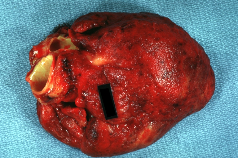
Tuberculous pericarditis: Gross, natural color, shaggy hemorrhagic exudate. This case is much more hemorrhagic than the typical tuberculous pericarditis.

Microscopic Pathology Images
-
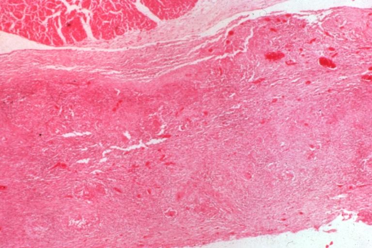
Tuberculous pericarditis.
-
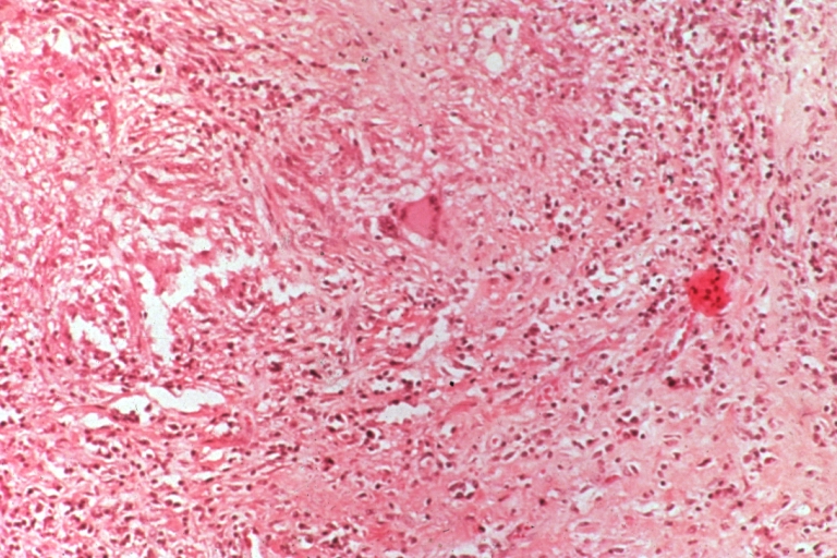
Tuberculous pericarditis.
-
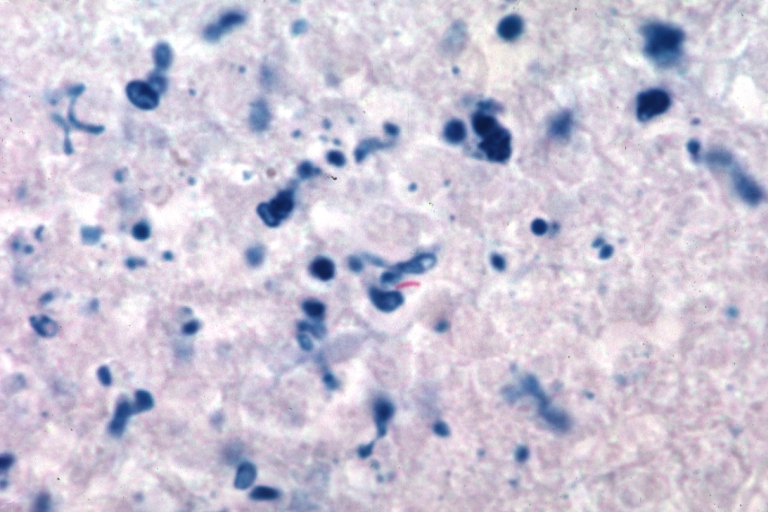
Tuberculous pericarditis: Micro oil acid fast stain. The organism easily seen.


-
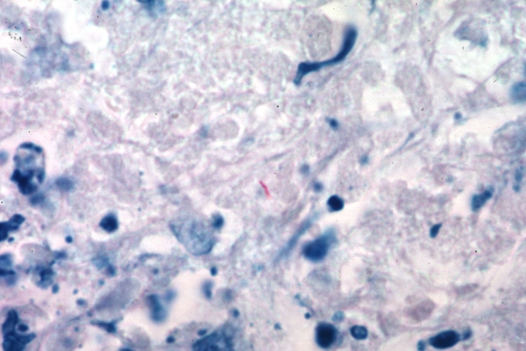
Tuberculous pericarditis: Micro oil acid fast stain. The organism easily seen.
-
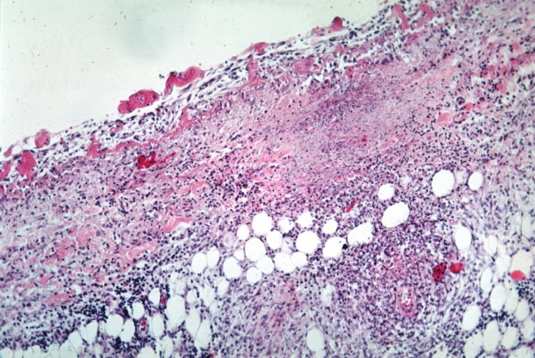
Tuberculous pericarditis: Micro med mag, H&E, a typical lesion.


References
- ↑ Peel AA (1948). "TUBERCULOUS PERICARDITIS". Br Heart J. 10 (3): 195–207. PMC 481044. PMID 18610109.
- ↑ Permanyer-Miralda G, Sagristá-Sauleda J, Soler-Soler J (1985). "Primary acute pericardial disease: a prospective series of 231 consecutive patients". Am J Cardiol. 56 (10): 623–30. PMID 4050698.
- ↑ Mayosi BM, Burgess LJ, Doubell AF (2005). "Tuberculous pericarditis". Circulation. 112 (23): 3608–16. doi:10.1161/CIRCULATIONAHA.105.543066. PMID 16330703.
- ↑ Sagristà-Sauleda J, Angel J, Sánchez A, Permanyer-Miralda G, Soler-Soler J (2004). "Effusive-constrictive pericarditis". N Engl J Med. 350 (5): 469–75. doi:10.1056/NEJMoa035630. PMID 14749455.