Subclavian vein
| Cardiology Network |
 Discuss Subclavian vein further in the WikiDoc Cardiology Network |
| Adult Congenital |
|---|
| Biomarkers |
| Cardiac Rehabilitation |
| Congestive Heart Failure |
| CT Angiography |
| Echocardiography |
| Electrophysiology |
| Cardiology General |
| Genetics |
| Health Economics |
| Hypertension |
| Interventional Cardiology |
| MRI |
| Nuclear Cardiology |
| Peripheral Arterial Disease |
| Prevention |
| Public Policy |
| Pulmonary Embolism |
| Stable Angina |
| Valvular Heart Disease |
| Vascular Medicine |
Editor-In-Chief: C. Michael Gibson, M.S., M.D. [1]
Overview
In human anatomy, the subclavian veins are two large veins, one on either side of the body. Its diameter is approximately that of a man's small finger. It is divided into right and left subclavian vein.
Path
Each subclavian vein is a continuation of the axillary vein and runs from the outer border of the first rib to the medial border of anterior scalene muscle.
From here it joins with the internal jugular vein to form the brachiocephalic vein (also known as "innominate vein"). The angle of union is termed the venous angle.
The subclavian vein follows the subclavian artery and is separated posteriorly by the insertion of anterior scalene.
Lymph
The thoracic duct drains into the left subclavian vein, near its junction with the left internal jugular vein.
It carries lymph (water and solutes) from the lymphatic system, as well as chylomicrons or chyle, formed in the intestines from dietery fat and lipids.
Etymology
The term subclavian can be broken down to: sub= below, and clavian= pertaining to the clavicle.
See also
Additional images
-
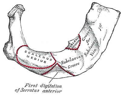
Peculiar ribs.
-
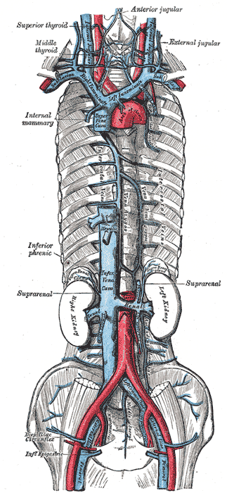
The venæ cavæ and azygos veins, with their tributaries.
-
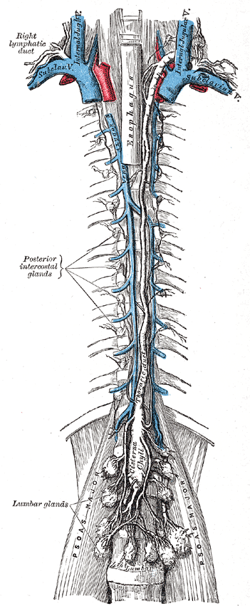
The thoracic and right lymphatic ducts.
-
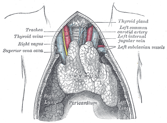
The thymus of a full-time fetus, exposed in situ.


