Help
Uploads by Gunnam
Jump to navigation
Jump to search
This special page shows all uploaded files.
File list
Items per page:
20
50
100
250
500
Username:
Include old versions of files
Go
First page
Previous page
Next page
Last page
Date
Name
Thumbnail
Size
Description
Versions
15:35, 3 January 2020
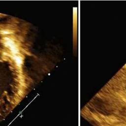
Transthoracic echocardiogram.jpg
(
file
)
8 KB
1
16:35, 20 February 2019
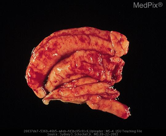
Thyroid pathology.jpg
(
file
)
8 KB
1
17:07, 8 February 2019
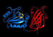
PTEN Gene.jpg
(
file
)
13 KB
1
15:50, 13 March 2020
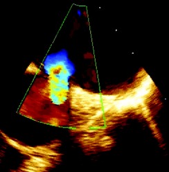
Patent ductus arteriosus (PDA) in Transesophageal echocardiography.jpg
(
file
)
16 KB
1
16:56, 20 February 2019
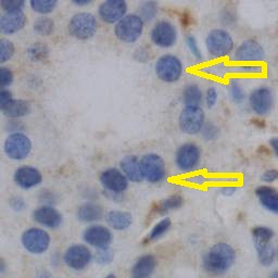
Immunohistochemistry in thyroid pathology.jpg
(
file
)
16 KB
1
20:01, 13 June 2019
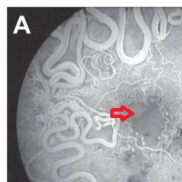
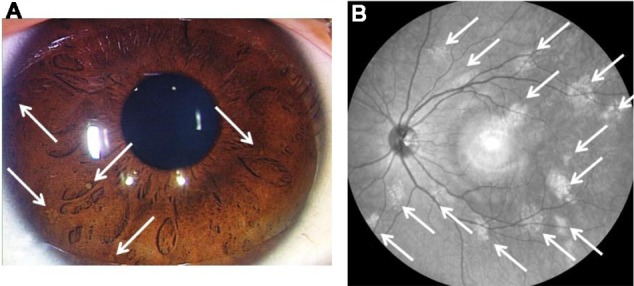
Macular ischemia and extensive arteriovenous communications and dilated intertwined vessels.jpg
(
file
)
19 KB
1
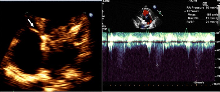
20:43, 22 April 2020
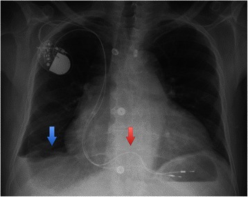
Pleural effusion in TR.jpg
(
file
)
23 KB
1
21:55, 12 March 2020
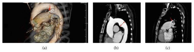
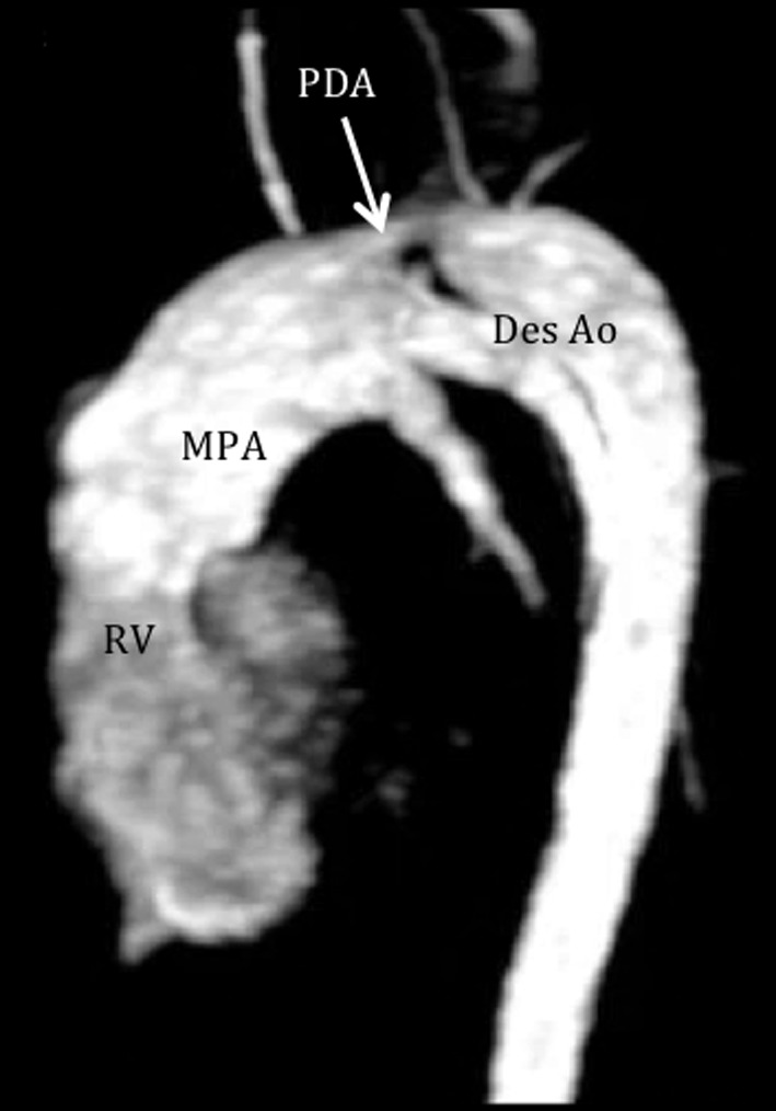
CT image showing the PDA.jpg
(
file
)
24 KB
1
16:47, 26 November 2019
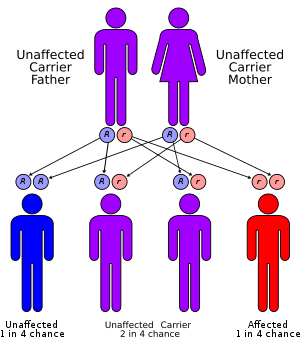
Autosomal recessive gene.png
(
file
)
24 KB
1
19:40, 23 January 2020
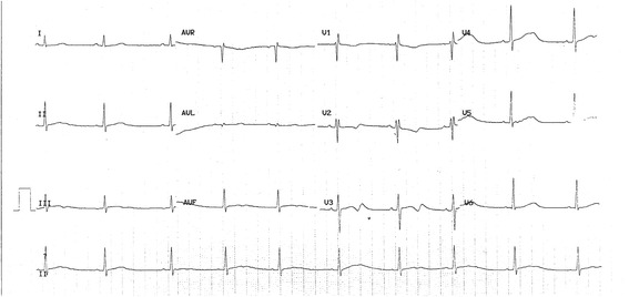
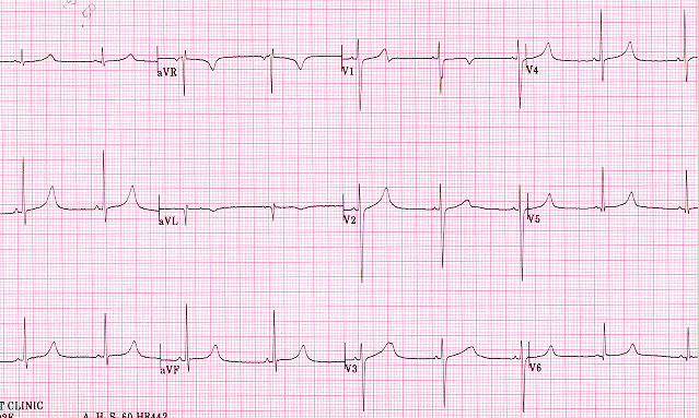
ECG in long QT syndrome.jpg
(
file
)
26 KB
1
16:37, 2 July 2019
Life According to Sam.jpg
(
file
)
27 KB
1
14:47, 26 April 2019
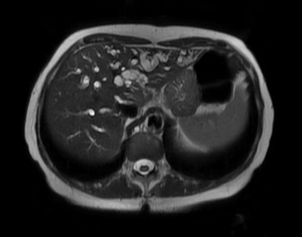
Lhermitte Duclos Disease.jpg
(
file
)
29 KB
1
19:46, 7 March 2018
Gallbladder cancer.jpg
(
file
)
32 KB
2
15:12, 14 September 2019
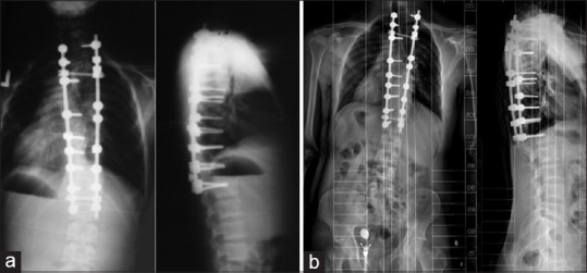
Aarskog-Scott syndrome X-ray.jpg
(
file
)
32 KB
1
14:36, 25 March 2020
Tricuspid annulus.jpg
(
file
)
34 KB
1
15:22, 7 February 2018
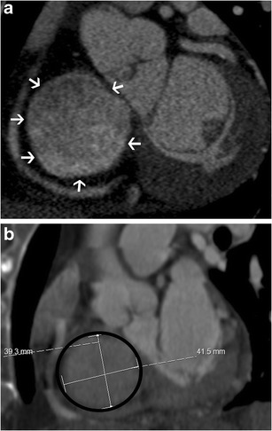
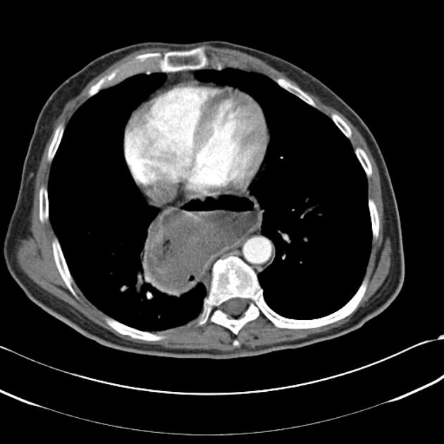
Ct ht.jpg
(
file
)
37 KB
1
14:28, 13 March 2020
Transesophageal echocardiogram of a patent ductus arteriosus.jpg
(
file
)
39 KB
1
17:26, 25 March 2020
Tricuspid stenosis in CT and MRI.jpg
(
file
)
40 KB
1
20:08, 9 April 2018
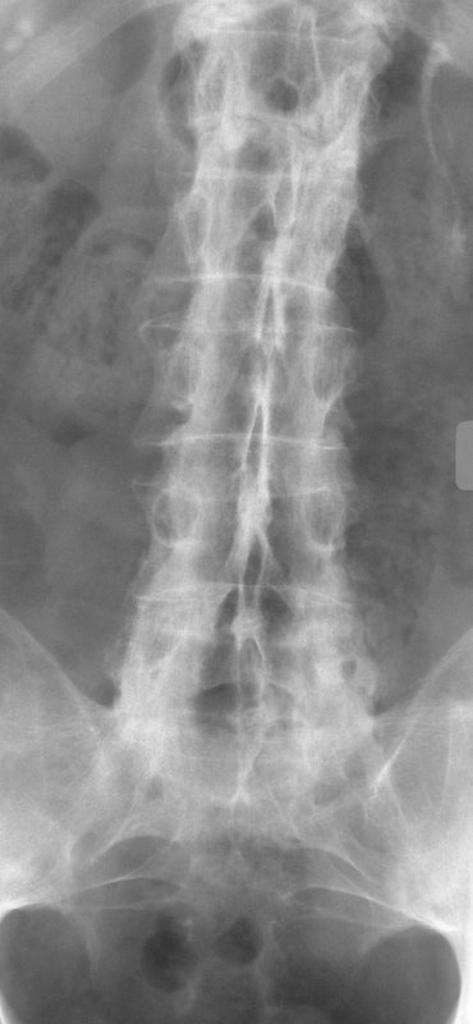
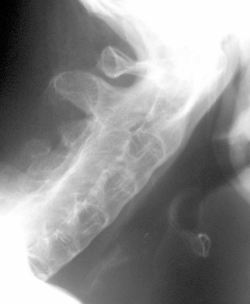
Ankylosing spondylitis DAGGER SPINE.jpg
(
file
)
43 KB
1
14:17, 13 September 2019
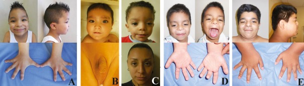
Aarskog syndrome.jpg
(
file
)
43 KB
1
19:34, 7 March 2018
Gallbladder-adenocarcinoma.jpg
(
file
)
44 KB
1
13:25, 26 September 2019
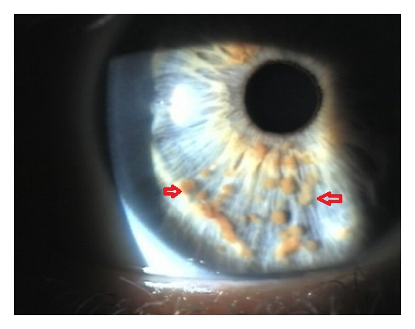
Lisch Nodule.jpg
(
file
)
46 KB
1
17:54, 8 February 2019
Lipoma.jpg
(
file
)
50 KB
1
21:18, 26 July 2018
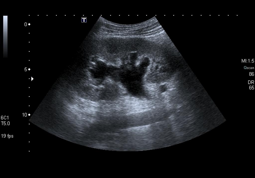
Kidney Stones On Ultrasound.jpg
(
file
)
55 KB
1
15:03, 13 April 2021
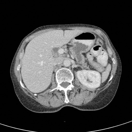
Pancreatic fistula CT.jpg
(
file
)
56 KB
1
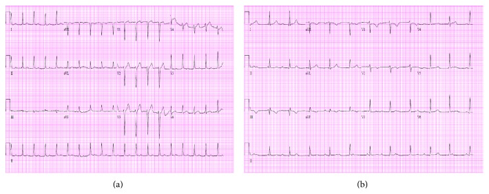
14:42, 4 March 2020
First-degree heart block and complete heart block.jpg
(
file
)
60 KB
1
13:56, 19 March 2020
Normal anatomy of tricuspid valve.jpg
(
file
)
61 KB
1
17:17, 8 February 2019
BRRS.jpg
(
file
)
62 KB
1
17:57, 7 August 2019
Acro-osteolysis.jpg
(
file
)
63 KB
1
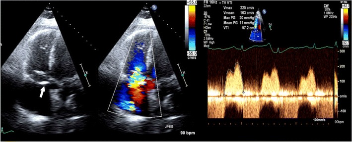
16:49, 19 March 2020
Tricuspid stenosis in echocardiography.jpg
(
file
)
65 KB
1
19:03, 26 July 2018
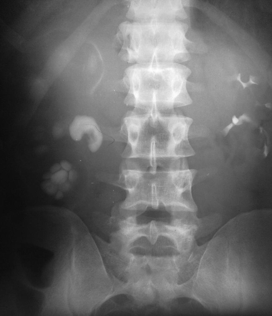
Severe-Hydronephrosis.jpg
(
file
)
65 KB
1
16:47, 19 March 2020
Normal echocardiographic appearance of tricuspid valve.jpg
(
file
)
65 KB
1
17:42, 13 February 2020
Pseudo “Tee - Pee sign”.jpg
(
file
)
66 KB
1
20:36, 22 April 2020
CMR four-chamber cine view. Seen here is the grossly dilated right heart, with an atrialized RV and dilated tricuspid annulus.jpg
(
file
)
68 KB
1
01:33, 23 April 2020
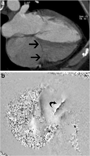
MRI velocity-encoding magnitude.jpg
(
file
)
68 KB
1
18:01, 10 April 2018
Ankylosing spondylitis.jpg
(
file
)
70 KB
1
16:11, 24 January 2019
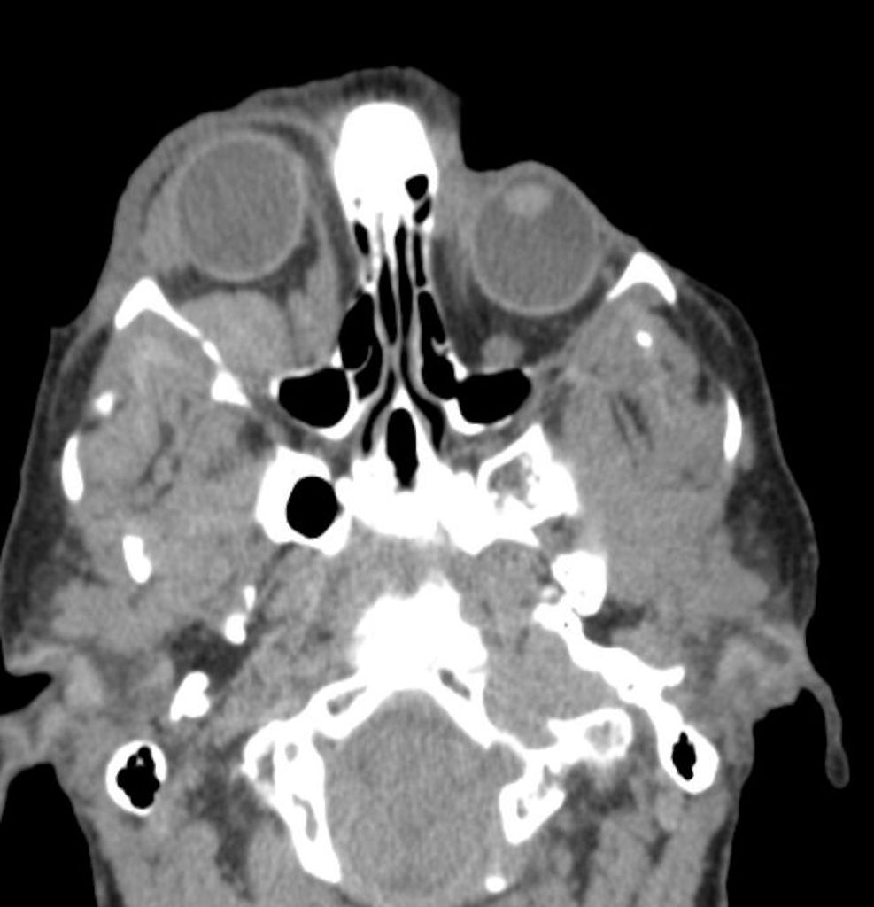
Head CT in BRBNS.jpg
(
file
)
71 KB
1
17:24, 4 December 2018
BRNS.jpg
(
file
)
72 KB
1
21:38, 26 July 2018
Rim-Sigh.jpg
(
file
)
73 KB
1
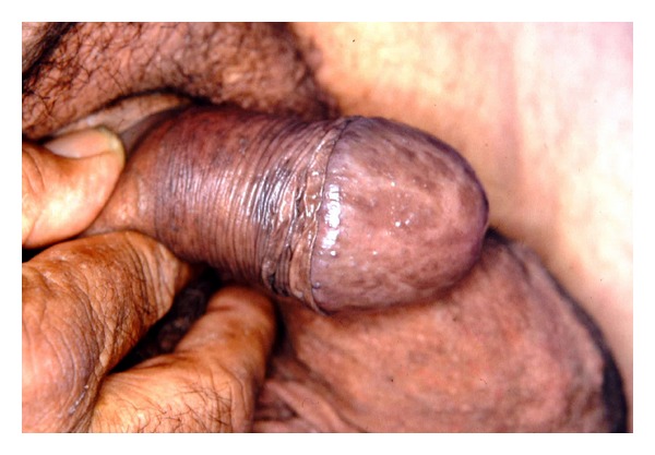
14:05, 11 February 2019
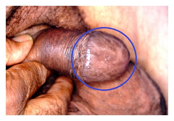
Penile lentigines in BRRS .jpg
(
file
)
76 KB
1
19:30, 7 March 2018
Gallbladder-adenocarcinoma-1.jpg
(
file
)
76 KB
1
13:30, 26 September 2019
Lisch nodules.jpg
(
file
)
78 KB
1
15:29, 31 December 2019
Romano-Ward syndrome .jpg
(
file
)
78 KB
1
18:18, 22 January 2019
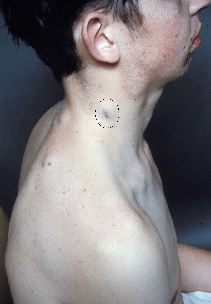
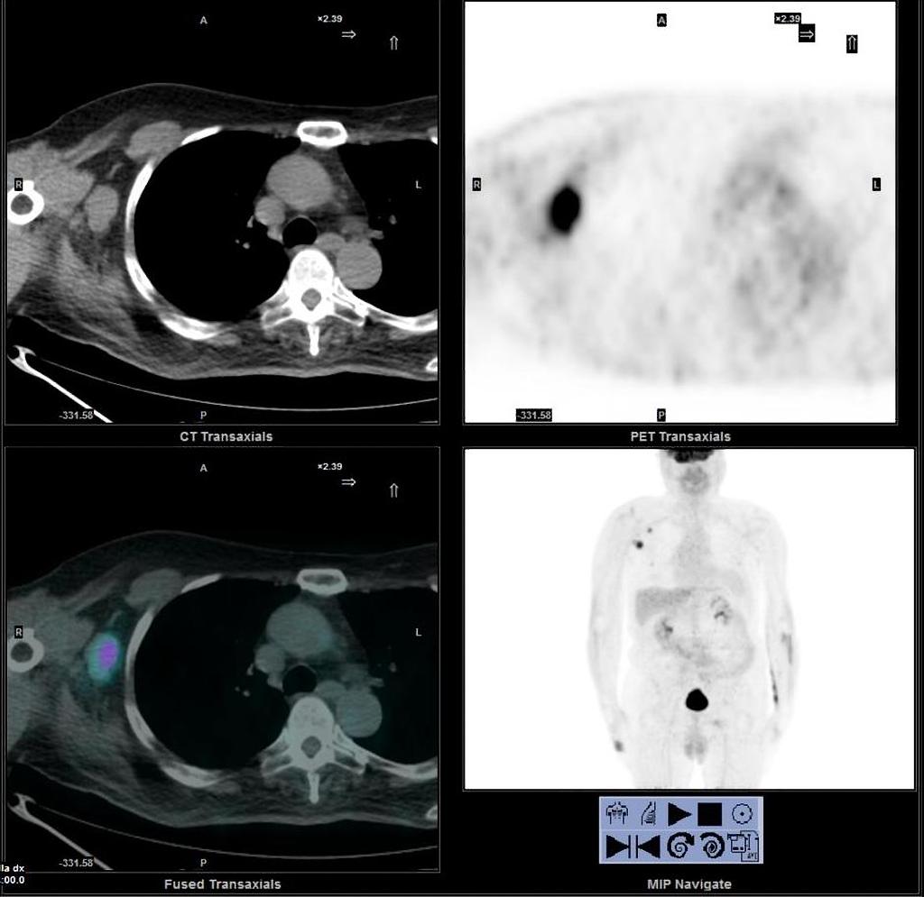
Metastatic-merkel-cell-carcinoma.jpg
(
file
)
79 KB
1
01:39, 6 March 2020
Multislice CT scan.jpg
(
file
)
79 KB
1
13:30, 13 March 2020
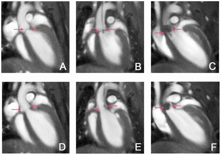
MRI demonstrates a patent ductus arteriosus.jpg
(
file
)
80 KB
1
16:50, 26 February 2020
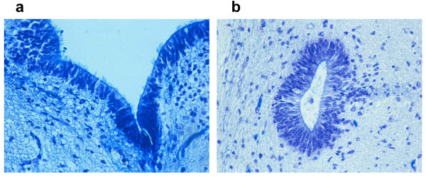
Pseudostratified ependymal layer.jpg
(
file
)
81 KB
1
01:15, 6 March 2020
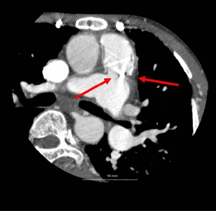
Vegetations on the aortic valve.jpg
(
file
)
83 KB
1
20:01, 9 April 2018
Ankylosing spondylitis Bamboo spine.jpg
(
file
)
84 KB
1
04:07, 15 April 2020
Lancisi sign.jpg
(
file
)
86 KB
1
First page
Previous page
Next page
Last page
Cookies help us deliver our services. By using our services, you agree to our use of cookies.
OK
Navigation menu
Personal tools
Log in
Request account
Namespaces
Special page
English
Views
More
Tools
User contributions
Logs
View user groups
Special pages
Printable version


_in_Transesophageal_echocardiography.jpg)