Uploads by Aditya Ganti
Jump to navigation
Jump to search
This special page shows all uploaded files.
| Date | Name | Thumbnail | Size | Description | Versions |
|---|---|---|---|---|---|
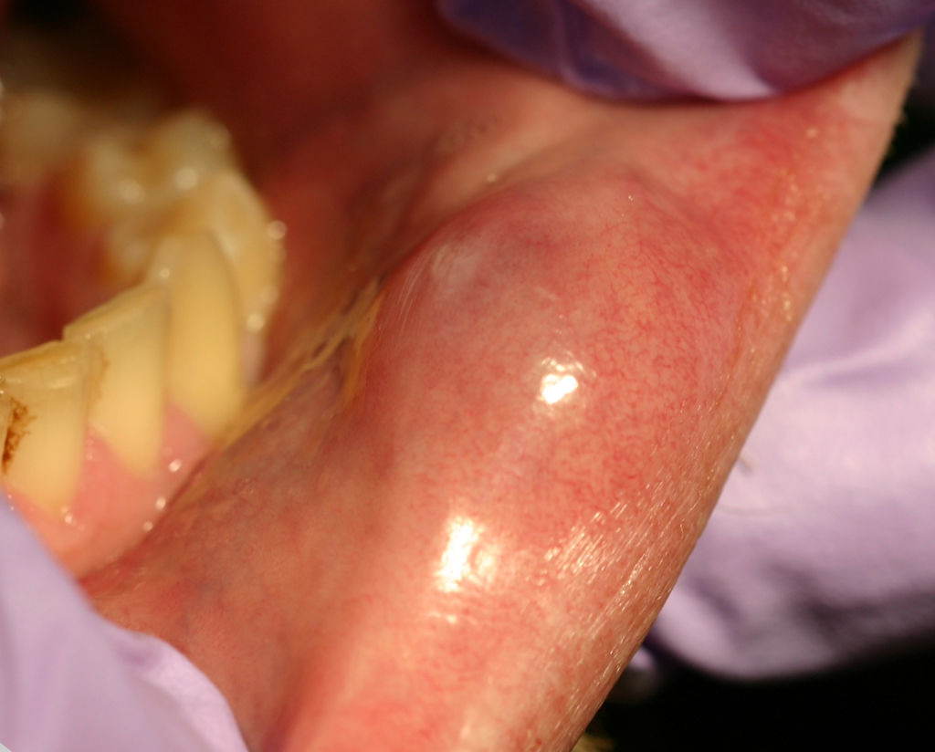
| 22:57, 12 February 2019 | 1024px-Mucocele02-17-06cropped.jpg (file) |  |
77 KB | 1 | |
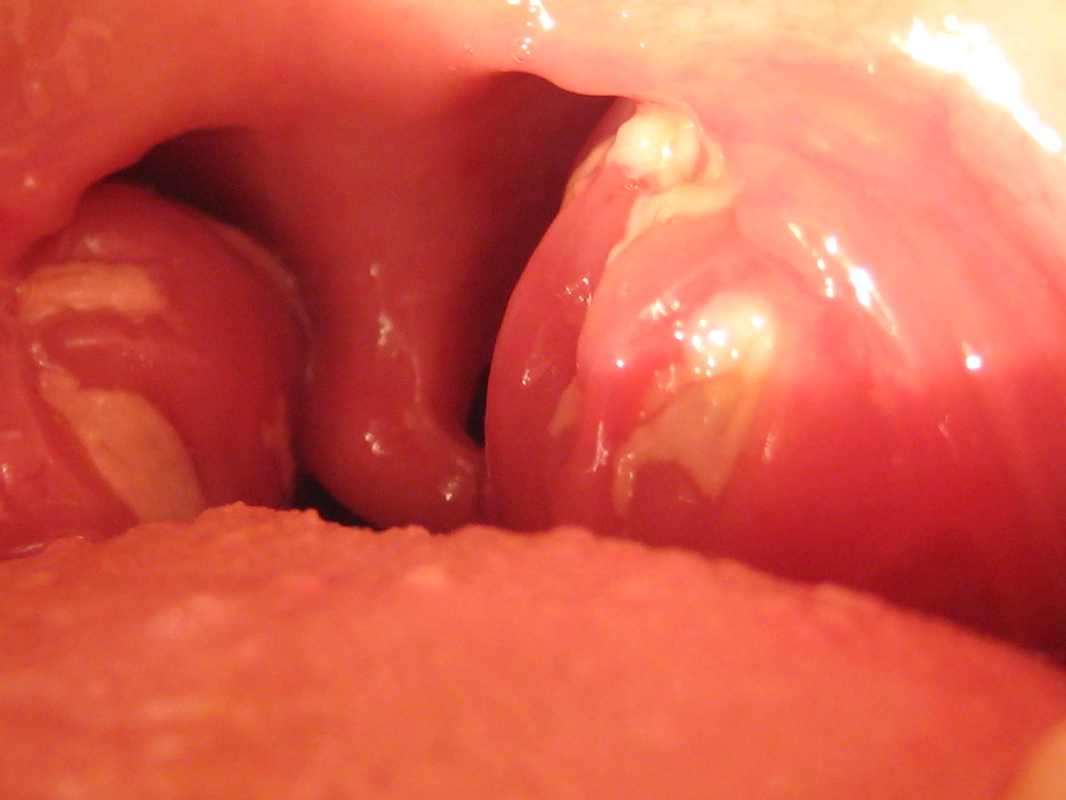
| 18:12, 12 February 2019 | 1200px-Mono tonsils.JPG (file) |  |
88 KB | 1 | |
| 18:56, 5 October 2017 | 7803935 orig.jpg (file) |  |
77 KB | 1 | |
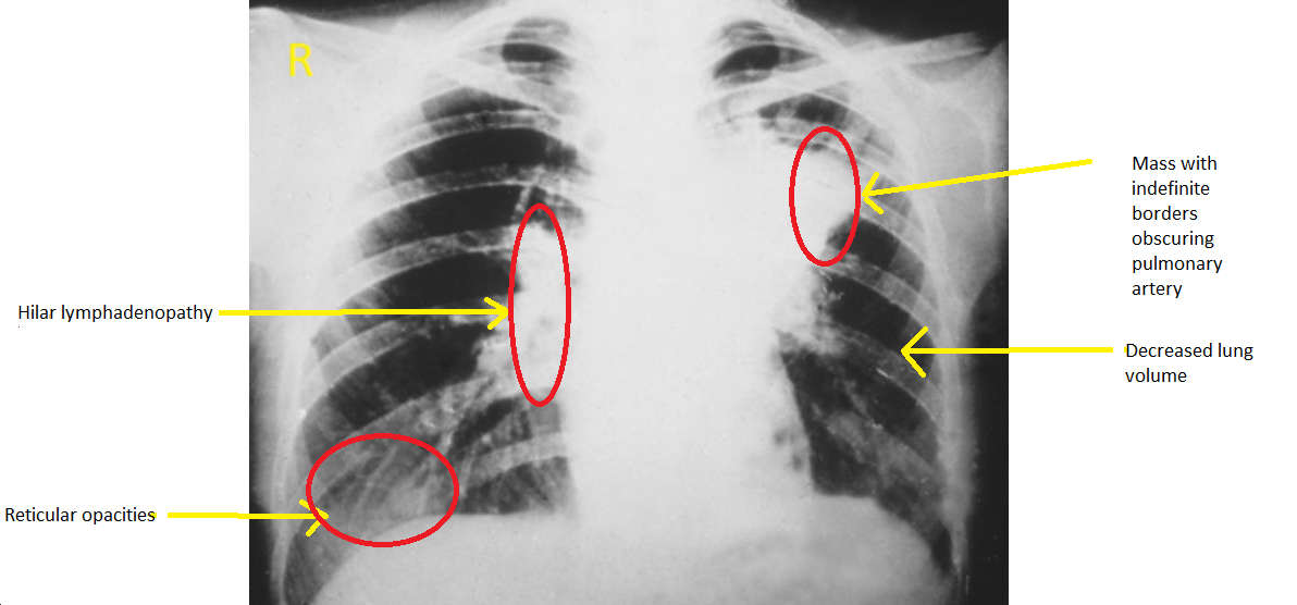
| 16:00, 29 October 2019 | ALL-Mediastinal Lymphadenopathy.gif (file) |  |
1.72 MB | 1 | |
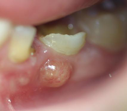
| 23:47, 12 February 2019 | Abces parulique.jpg (file) |  |
74 KB | 1 | |
| 18:30, 16 March 2017 | Actinomyces.high magnification.jpg (file) |  |
97 KB | 2 | |
| 16:05, 21 March 2017 | Actinomycosis1.jpg (file) |  |
39 KB | 1 | |
| 17:30, 9 January 2018 | Acute-appendicitis-paediatric-2.jpg (file) |  |
89 KB | 2 | |
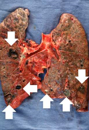
| 18:08, 6 February 2017 | Acute lung abscess.png (file) |  |
369 KB | 1 | |
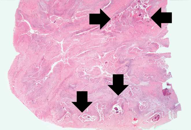
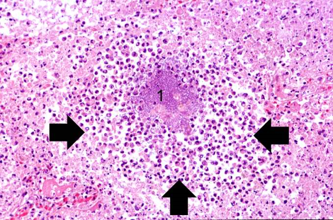
| 17:36, 6 February 2017 | Acute lung abscess microscopic.png (file) |  |
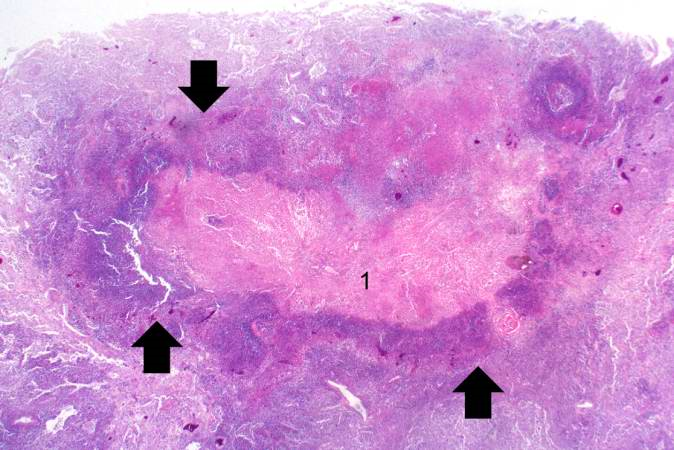
855 KB | A high-power photomicrograph of the lung demonstrating a small abscess full of inflammatory cells (primarily neutrophils) (arrows). 1.There is a bacterial colony in the center of this abscess | 1 |
| 17:24, 21 September 2017 | Addisons CT.gif (file) |  |
161 KB | 1 | |
| 15:56, 21 September 2017 | Addisons hyperpigmentation.jpg (file) |  |
5 KB | 1 | |
| 19:56, 19 January 2017 | Aditya Ganti.jpg (file) |  |
133 KB | 1 | |
| 22:12, 13 December 2017 | Age wise incidence.png (file) |  |
52 KB | 1 | |
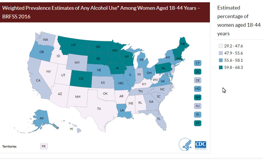
| 18:03, 8 September 2018 | Alcohol.png (file) |  |
105 KB | 1 | |
| 14:48, 21 September 2017 | Alzheimers-disease-pet-ct-1.jpg (file) |  |
67 KB | 1 | |
| 14:54, 21 September 2017 | Alzheimers-disease-pet-ct-jpg.jpeg (file) |  |
36 KB | 1 | |
| 16:28, 10 April 2018 | Ankylosing-spondylitis-and-chalk-fracture.jpg (file) |  |
78 KB | 1 | |
| 18:04, 9 January 2018 | Appendicitis-gross-pathology.png (file) |  |
368 KB | 1 | |
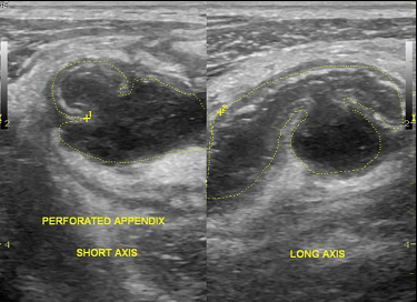
| 17:34, 9 January 2018 | Appendicitis-perforated.png (file) |  |
172 KB | 4 | |
| 17:25, 22 February 2017 | Appendicular abscess CT gif.gif (file) |  |
2.11 MB | 2 | |
| 22:00, 22 February 2017 | Appendicular abscess USG.gif.gif (file) |  |
263 KB | 1 | |
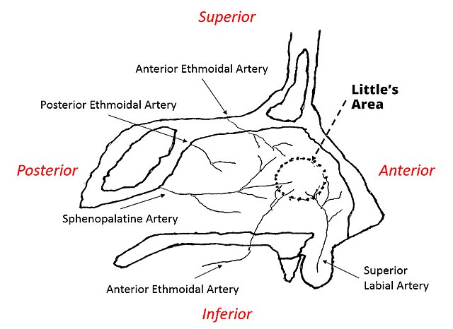
| 10:56, 30 March 2021 | Arteries of the Nose.jpg (file) |  |
48 KB | 2 | |
| 17:08, 7 March 2017 | Blastomycosis Chest Xray.png (file) |  |
353 KB | 1 | |
| 13:14, 9 March 2017 | Blastomycosis Chest Xray1.png (file) |  |
248 KB | 1 | |
| 15:03, 13 July 2017 | Bourbon virus.gif (file) | 20 KB | 1 | ||
| 15:14, 13 July 2017 | Bourbon virus sphere (EID 2015 Fig 2b).jpg (file) | .jpg) |
73 KB | 1 | |
| 02:58, 18 April 2017 | CT spine.gif.gif (file) |  |
1.08 MB | 1 | |
| 17:42, 6 February 2017 | Chronic lung abscess microscopic.png (file) |  |
779 KB | The center of the abscess contains necrotic debris (1) and there is a rim of viable inflammatory cells (arrows) surrounding this abscess. | 1 |
| 17:34, 20 November 2017 | Colonic blood supply1.gif (file) |  |
79 KB | 1 | |
| 00:10, 18 February 2019 | Condyloma acuminatum.jpg (file) |  |
43 KB | 1 | |
| 17:58, 29 June 2020 | Covid-19-jpg.jpeg (file) |  |
224 KB | 1 | |
| 17:51, 29 June 2020 | Covid-19-pneumonia-20.jpg (file) |  |
130 KB | 1 | |
| 03:44, 25 June 2020 | Covid-5.jpeg (file) |  |
118 KB | 1 | |
| 02:22, 16 July 2020 | Covid-postinflammatory.jpeg (file) |  |
51 KB | 1 | |
| 19:46, 20 December 2017 | Crohn's transmural path 1.png (file) | 212 KB | 1 | ||
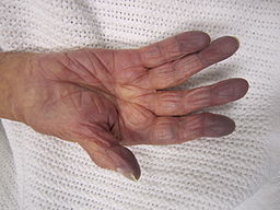
| 21:33, 6 March 2018 | Cynosis.JPG (file) |  |
13 KB | 1 | |
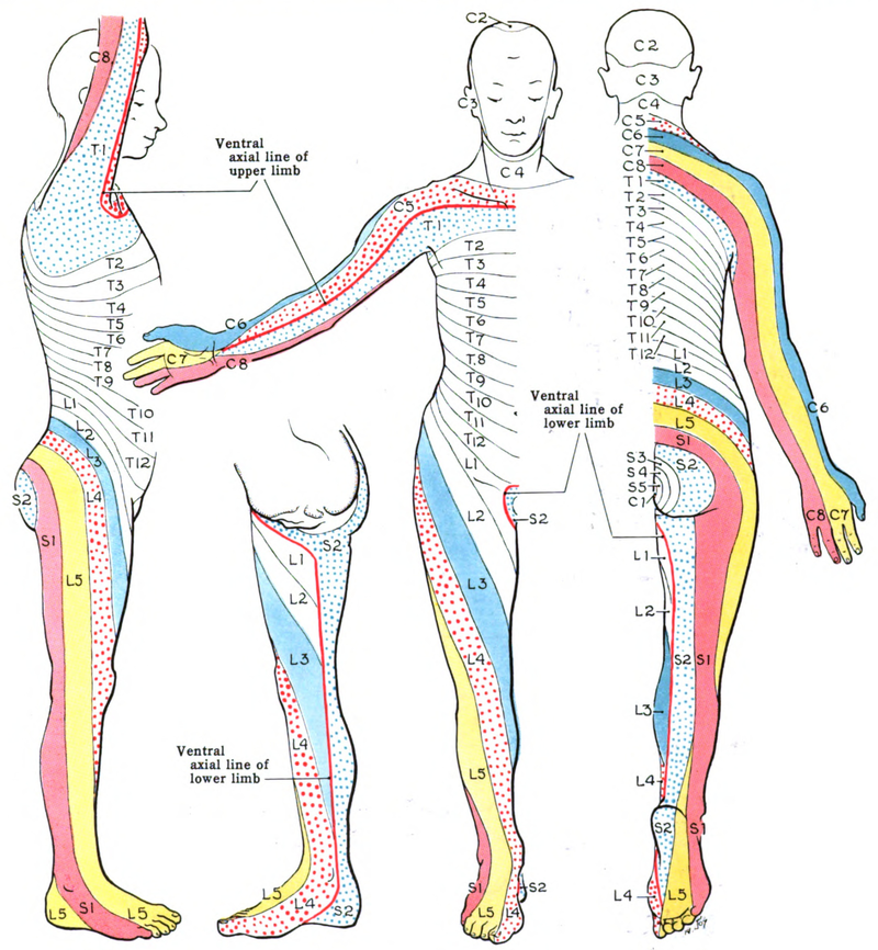
| 15:17, 13 April 2017 | Diagram of the spinal cord CRUK 046.svg.png (file) |  |
33 KB | 2 | |
| 14:46, 4 April 2017 | Diphtherial pharyngitis 1.jpg (file) |  |
205 KB | 1 | |
| 22:06, 6 November 2017 | Drug-induced hepat.gif (file) |  |
1.26 MB | 1 | |
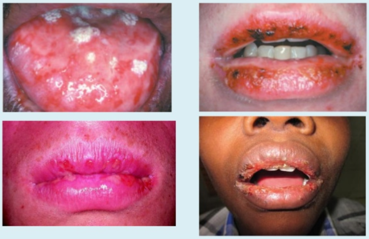
| 20:18, 12 February 2019 | EM oral.png (file) |  |
527 KB | 1 | |
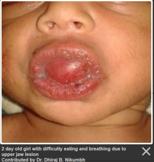
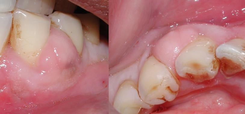
| 05:14, 13 February 2019 | Eplis.png (file) |  |
316 KB | 1 | |
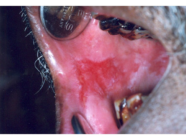
| 16:38, 11 February 2019 | Erythroplakia1.jpg (file) |  |
63 KB | 1 | |
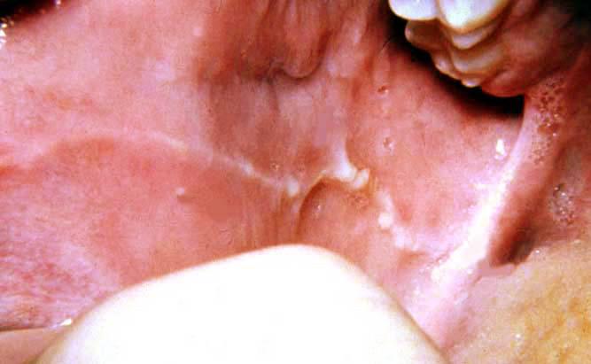
| 16:25, 11 February 2019 | Frictional hyperkeratosis.jpg (file) |  |
27 KB | 1 | |
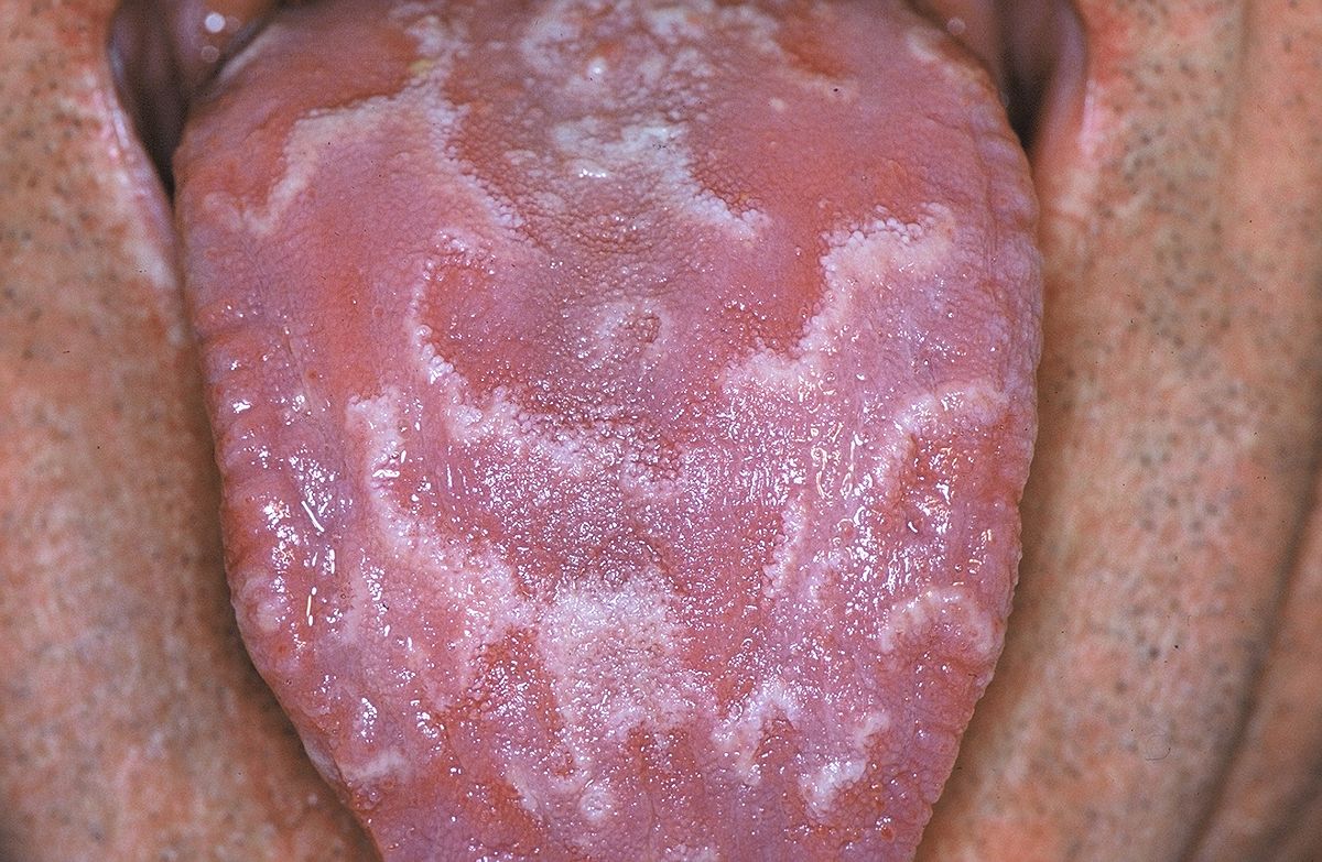
| 15:09, 11 February 2019 | Geographic tongue 01.JPG (file) |  |
209 KB | 1 | |
| 23:12, 12 February 2019 | GingivalAbscess.jpg (file) |  |
27 KB | 1 | |
| 16:03, 13 April 2017 | Grant 1962 663.png (file) |  |
776 KB | 1 | |
| 18:56, 9 August 2017 | Gynecomastia 2 (1).jpg (file) | .jpg) |
468 KB | 1 | |
| 18:59, 9 August 2017 | Gynecomastia 2 (2).jpg (file) | .jpg) |
124 KB | 1 | |
| 19:21, 27 June 2017 | HAT ga 2014 490.png (file) |  |
32 KB | 1 |