Uploads by Ssharfaei
Jump to navigation
Jump to search
This special page shows all uploaded files.
| Date | Name | Thumbnail | Size | Description | Versions |
|---|---|---|---|---|---|
| 23:39, 25 November 2017 | Rickets-16.png (file) |  |
521 KB | Rickets: Metaphyseal flaring and fraying seen at both proximal and distal tibia. There is slight outward bowing of both tibias. Case courtesy of Dr Henry Knipe, Radiopaedia.org, rID: 41684 https://radiopaedia.org/cases/41684 | 1 |
| 15:40, 22 November 2017 | LowKECG.png (file) |  |
1.24 MB | Hypokalemia | 1 |
| 21:37, 20 November 2017 | Anatomy-Male.ppt (file) | 285 KB | 2 | ||
| 21:33, 20 November 2017 | Anatomy-Female.ppt (file) | 770 KB | 2 | ||
| 16:03, 17 November 2017 | Whipple's disease1.gif (file) |  |
552 KB | 1 | |
| 15:57, 17 November 2017 | Whipple's disease.gif (file) |  |
552 KB | 3 | |
| 15:05, 16 November 2017 | Whipple disease Acid fast stain.jpg (file) |  |
847 KB | 1 | |
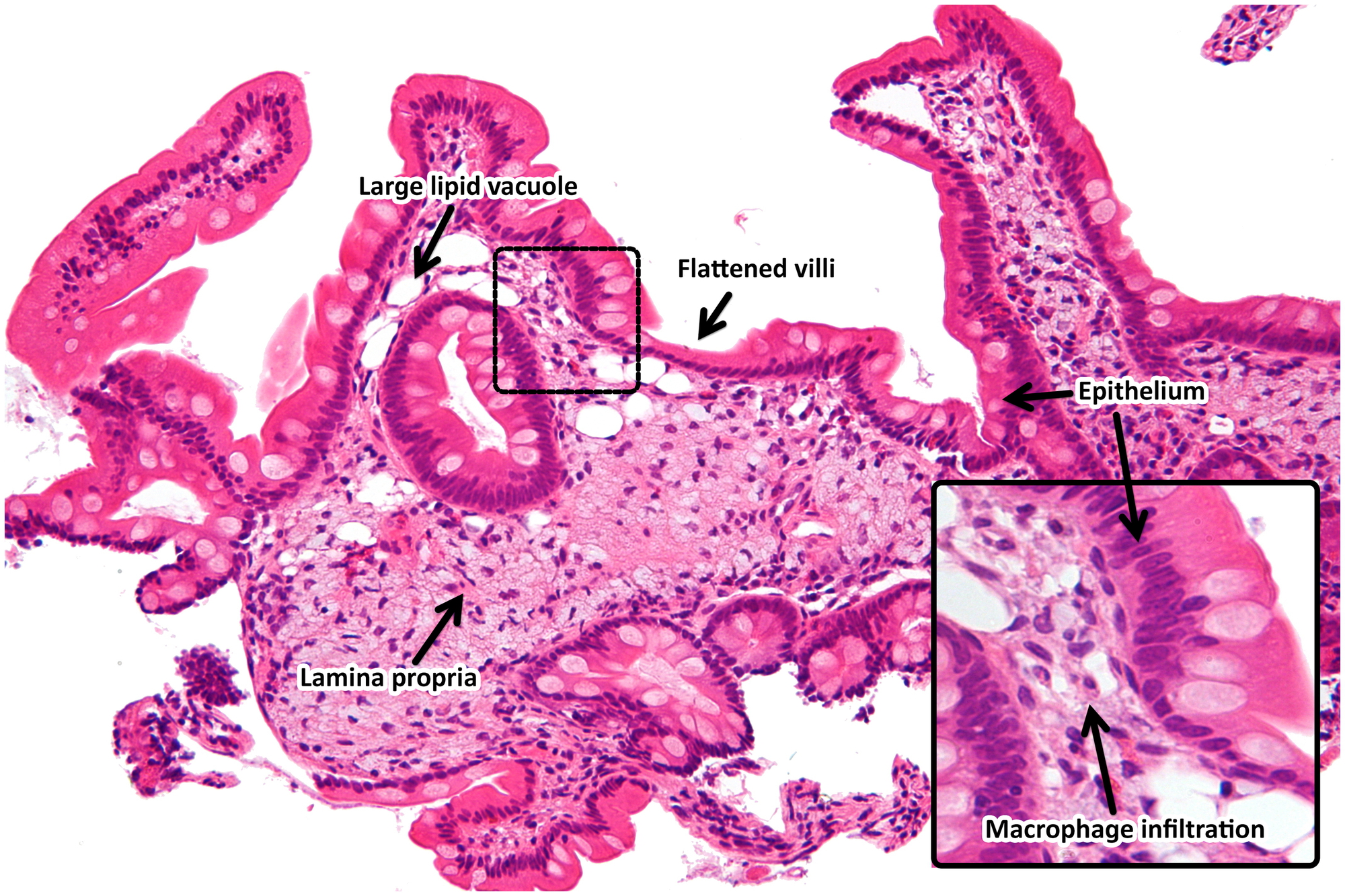
| 15:04, 16 November 2017 | Light microscopy of intestine-Whipples Disease.jpg .jpg (file) |  |
430 KB | 1 | |
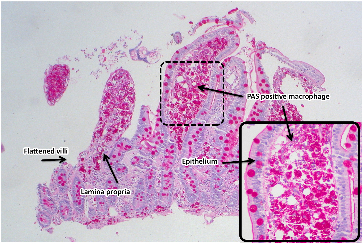
| 15:03, 16 November 2017 | Whipple disease PAS positive3.jpg (file) |  |
1.08 MB | 1 | |
| 15:02, 16 November 2017 | Whipple disease PAS positive2.jpg (file) |  |
252 KB | 1 | |
| 15:02, 16 November 2017 | Whipple disease PAS positive1.jpg (file) |  |
308 KB | 1 | |
| 15:01, 16 November 2017 | Whipple disease HE.jpg (file) |  |
1.96 MB | 1 | |
| 14:59, 16 November 2017 | Whipple disease GMS stain.jpg (file) |  |
1.45 MB | 1 | |
| 14:58, 16 November 2017 | Whipple disease PAS stain-m1.jpg (file) |  |
1.49 MB | 1 | |
| 15:32, 15 November 2017 | Whipple disease PAS stain 3.jpg (file) |  |
1.83 MB | 1 | |
| 15:32, 15 November 2017 | Whipple disease PAS stain 2.jpg (file) |  |
1.42 MB | 1 | |
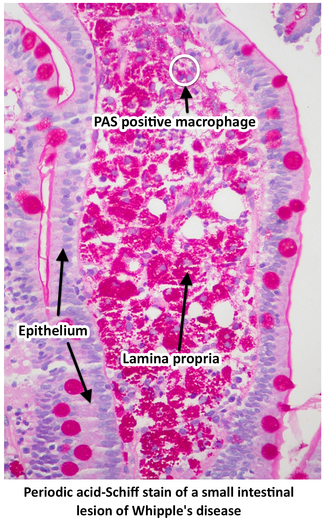
| 15:26, 15 November 2017 | Whipple disease PAS stain 1.jpg (file) |  |
1.48 MB | Periodic acid-Schiff stain of a small intestinal lesion of Whipple's disease by Ed Uthman from Houston, TX, USA - Whipple's Disease, PAS https://commons.wikimedia.org/w/index.php?curid=30104677 | 1 |
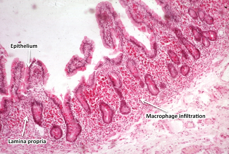
| 21:26, 13 November 2017 | Light microscopy of intestine-Whipples Disease.jpg (file) |  |
422 KB | light microscopy of intestine; Whipples Disease: Alcian blue with apparently eosin counterstain enlarged villus with many macrophages. From PEIR - University of Alabama at Birmingham Department of Pathology | 2 |
| 19:29, 9 November 2017 | Small bilateral pleural effusions.jpg (file) |  |
146 KB | 1 | |
| 16:05, 27 October 2017 | Tropheryma whipplei.jpeg (file) |  |
52 KB | 1 | |
| 19:44, 17 October 2017 | Whipple disease very high mag.jpg (file) |  |
308 KB | Very high magnification micrograph of Whipple's disease, also Whipple disease. H&E stain. Duodenal biopsy. The images show the characteristic feature of Whipple's disease; foamy macrophages are present in the lamina propria. | 1 |
| 19:43, 17 October 2017 | Whipple disease high mag.jpg (file) |  |
199 KB | High magnification micrograph of Whipple's disease, also Whipple disease. H&E stain. Duodenal biopsy. The images show the characteristic feature of Whipple's disease; foamy macrophages are present in the lamina propria. | 1 |
| 19:42, 17 October 2017 | Whipple disease - intermed mag.jpg (file) |  |
273 KB | Intermediate magnification micrograph of Whipple's disease, also Whipple disease. H&E stain. Duodenal biopsy. The images show the characteristic feature of Whipple's disease; foamy macrophages are present in the lamina propria. | 1 |
| 19:36, 17 October 2017 | Whipple disease low mag.jpg (file) |  |
1.52 MB | Low magnification micrograph of Whipple's disease. H&E stain. Duodenal biopsy. The images show the characteristic feature of Whipple's disease; foamy macrophages are present in the lamina propria. | 1 |
| 01:57, 16 May 2017 | SadafSharfaei.jpg (file) |  |
906 KB | 1 |