Uploads by Ssharfaei
Jump to navigation
Jump to search
This special page shows all uploaded files.
| Date | Name | Thumbnail | Size | Description | Versions |
|---|---|---|---|---|---|
| 14:32, 5 December 2017 | Crohn Jejunum.png (file) |  |
300 KB | Partial jejunum affected by morbus Crohn. Source: Wikimedia.org By Jaroslav Cehovsky - Camera, Public Domain, https://commons.wikimedia.org/w/index.php?curid=1458390 | 1 |
| 19:33, 3 April 2018 | Dermatomyositis5.jpg (file) |  |
399 KB | Dermatomyositis, Gottron's papules. Erythematous plaques overlying the elbows in two patients with juvenile dermatomyositis. In some patients, small erythematous plaques may overly the extensor aspects of larger joints, such as the elbows, knees, and m... | 1 |
| 16:09, 5 December 2017 | Diagram of the small bowel 01 CRUK 045.jpg (file) |  |
254 KB | Diagram of the small bowel 01. Source: Wikimedia.org By Cancer Research UK - Original email from CRUK, CC BY-SA 4.0, https://commons.wikimedia.org/w/index.php?curid=34332940 | 1 |
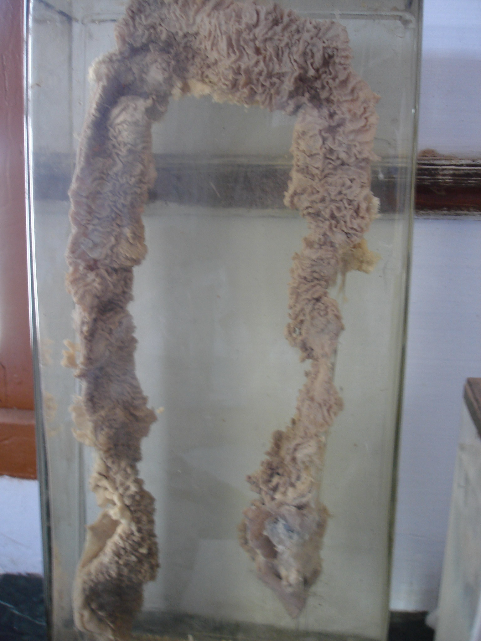
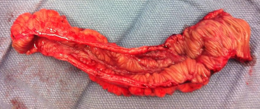
| 15:30, 30 January 2018 | Familial Adenomatous Polyposis intestine.jpg (file) |  |
497 KB | 2 | |
| 15:59, 30 January 2018 | Familial adenomatous polyposis.jpg (file) |  |
78 KB | 1 | |
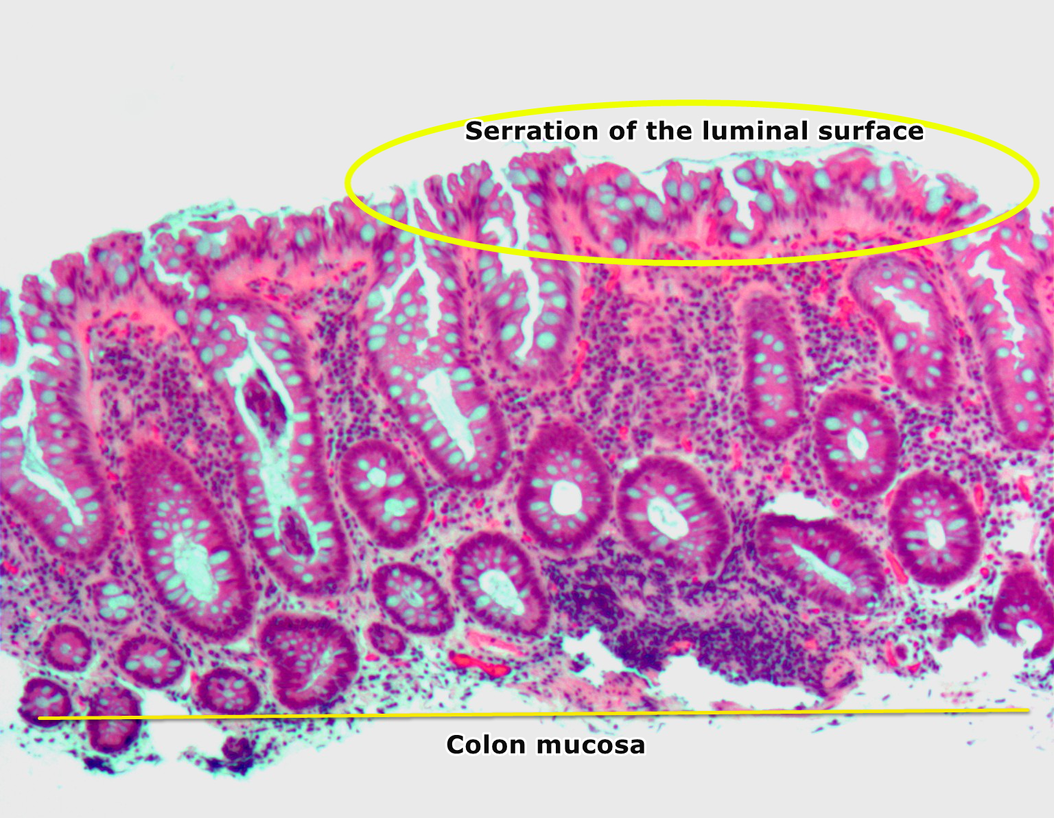
| 21:02, 26 January 2018 | Hyperplastic polyp2.jpg (file) |  |
946 KB | 3 | |
| 17:39, 23 January 2018 | Hyperplastic polyp of the colon, HE.png (file) |  |
1.93 MB | 1 | |
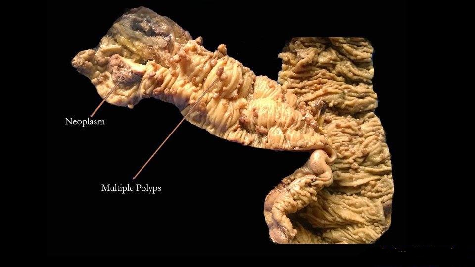
| 17:12, 20 December 2017 | Image of resected colon segment with cancer & 4 nearby polyps plus schematic of field defects with sub-clones.jpg (file) |  |
625 KB | 1 | |
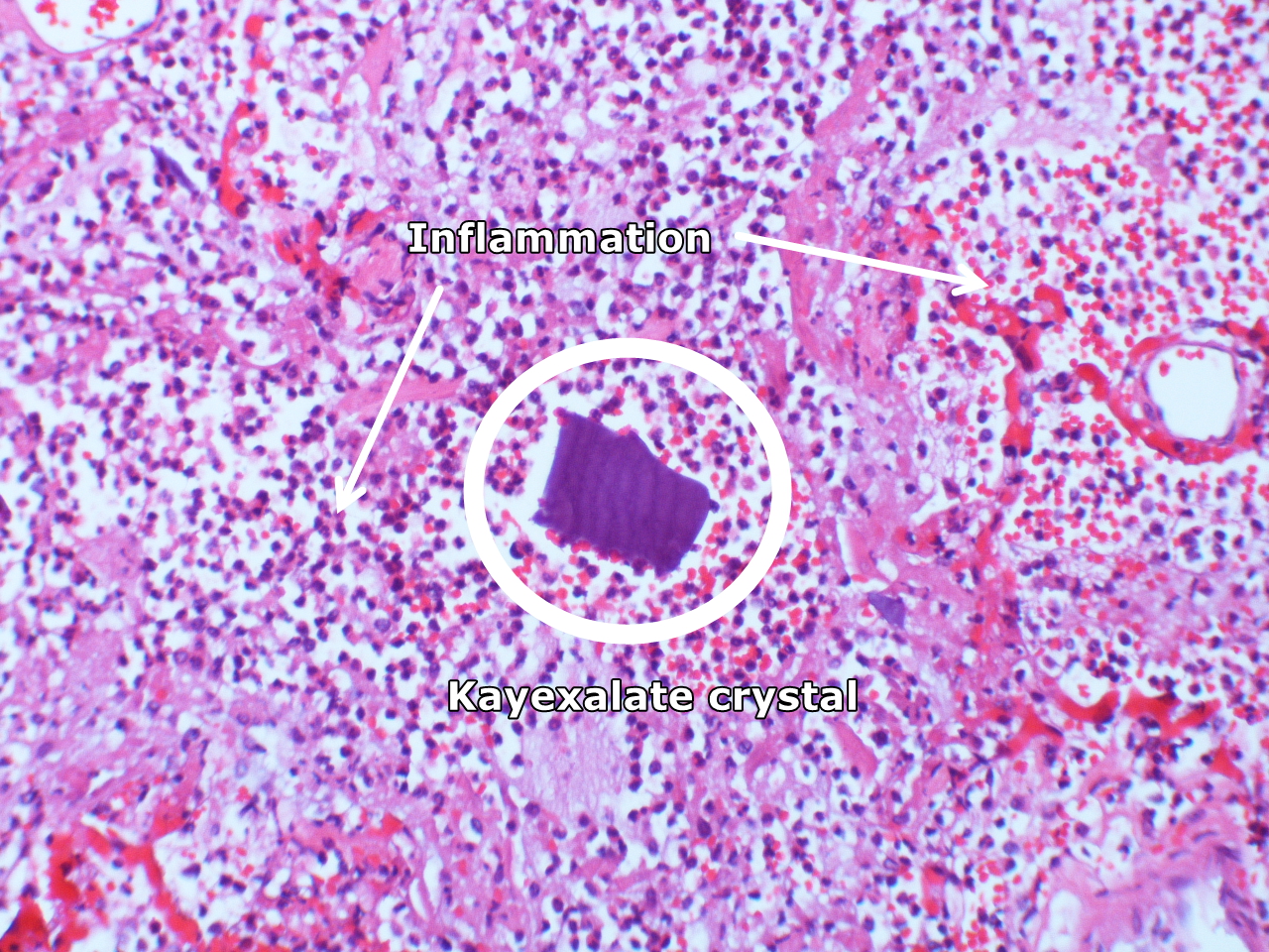
| 01:46, 31 March 2018 | Kayexalate aspiration Case 125 (4692318776).jpg (file) | .jpg) |
1.35 MB | Intraalveolar kayexalate crystal; acute pneumonitis. By Yale Rosen from USA - Kayexalate aspiration Case 125Uploaded by CFCF, CC BY-SA 2.0, Via Wikimedia<ref name="urlFile:Kayexalate aspiration Case 125 (4692318776).jpg - Wikimedia Commons">{{cite web... | 1 |
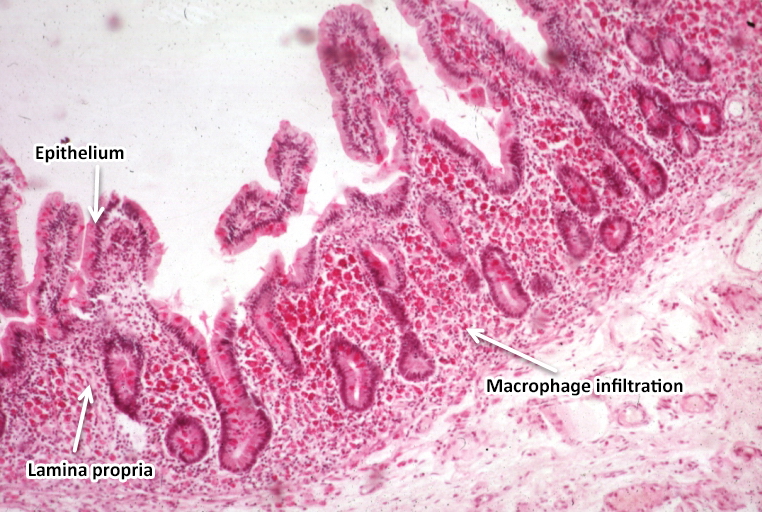
| 21:26, 13 November 2017 | Light microscopy of intestine-Whipples Disease.jpg (file) |  |
422 KB | light microscopy of intestine; Whipples Disease: Alcian blue with apparently eosin counterstain enlarged villus with many macrophages. From PEIR - University of Alabama at Birmingham Department of Pathology | 2 |
| 15:04, 16 November 2017 | Light microscopy of intestine-Whipples Disease.jpg .jpg (file) |  |
430 KB | 1 | |
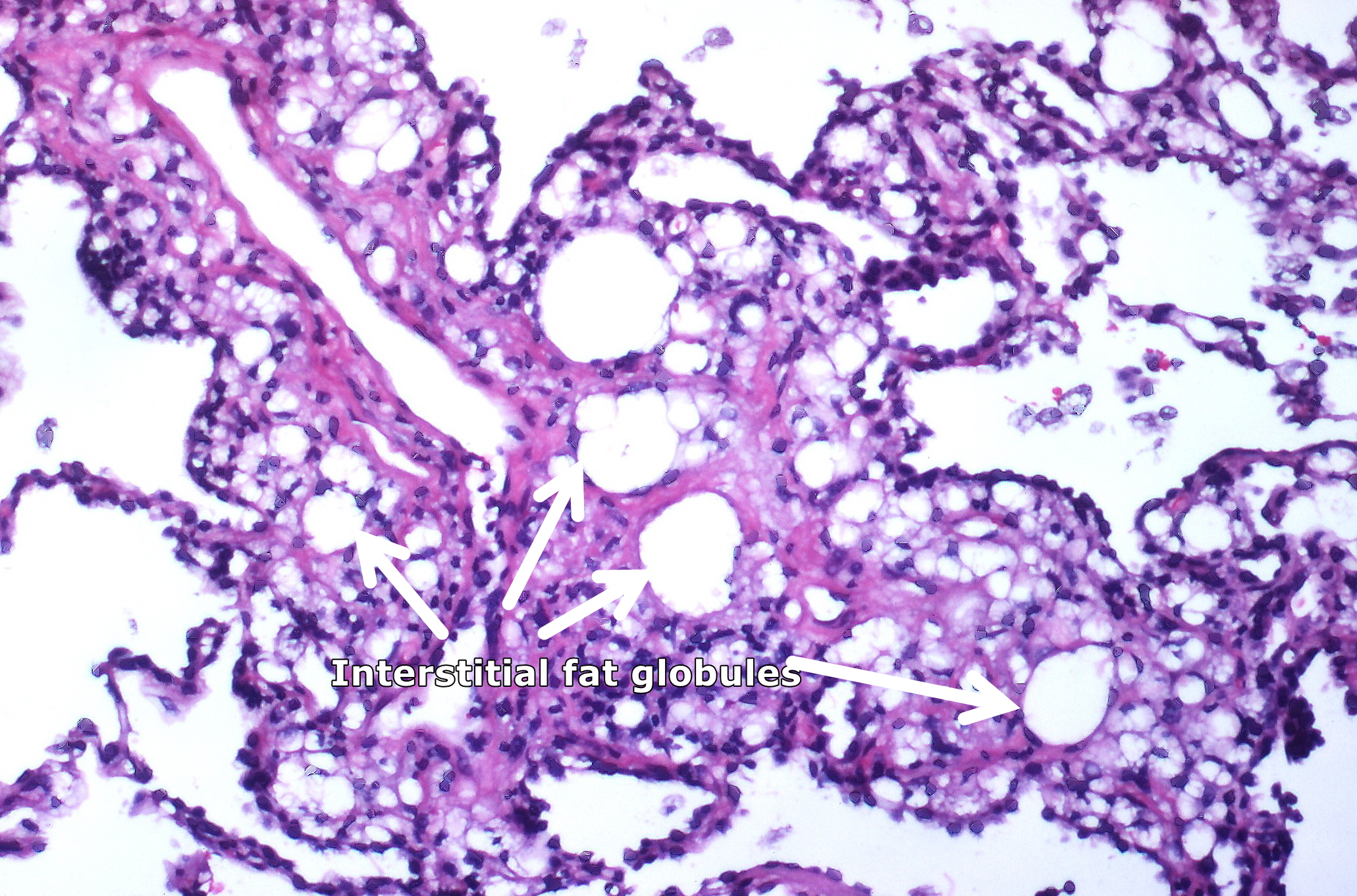
| 01:50, 31 March 2018 | Lipid pneumonia, exogenous (3791887936).jpg (file) | .jpg) |
1.81 MB | Numerous interstitial fat globules of varying size accompanied by inflammation and fibrosis is characterstic of chronic lipid pneumonia secondary to lipid aspiration. By Yale Rosen from USA - Lipid pneumonia, exogenousUploaded by CFCF, CC BY-SA 2.0, Vi... | 1 |
| 15:40, 22 November 2017 | LowKECG.png (file) |  |
1.24 MB | Hypokalemia | 1 |
| 19:46, 11 May 2018 | MediaWiki Vector skin action arrow.png (file) | 231 bytes | 1 | ||
| 16:57, 20 June 2018 | Narrative review-Template.pdf (file) | 211 KB | 1 | ||
| 17:59, 3 May 2018 | Neuropathology case XII 01.jpg (file) |  |
667 KB | Muscle biopsy frozen section specimen - Myositis (HE stain)-Partial invasion of lymphocytes within the muscle fibers via librepathology.org | 1 |
| 16:50, 13 March 2019 | Oesophageal-squamous-cell-carcinoma-2.jpg (file) |  |
243 KB | 2 | |
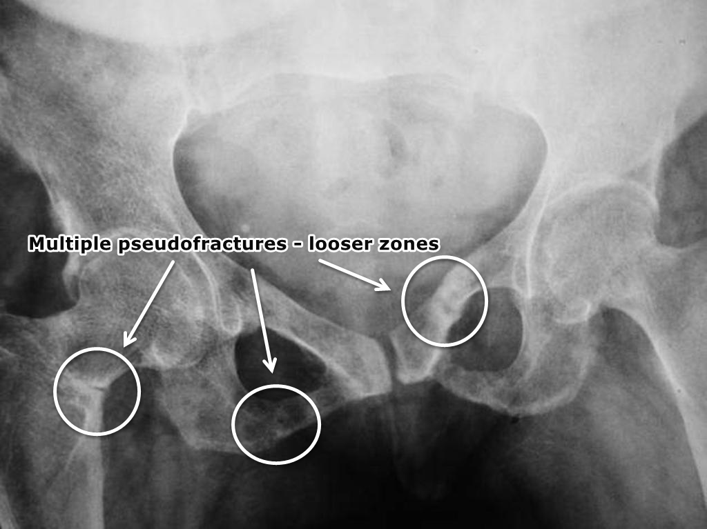
| 00:53, 26 November 2017 | Osteomalacia-looser-zones-1.png (file) |  |
452 KB | Osteopenic changes involving bony pelvis and proximal femurs. Multiple pseudofractures/Looser zones are seen involving superior and inferior pubic rami bilaterally. There is also a transcervical fracture on the right side. Case courtesy of Dr Iqbal Na... | 1 |
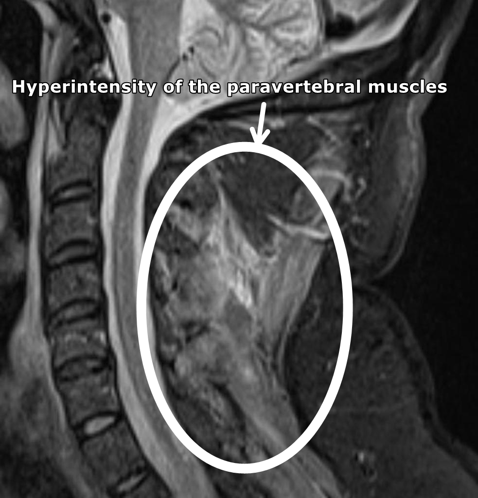
| 14:41, 13 April 2018 | Polymyositis-1.JPG (file) |  |
238 KB | MRI (sagittal STIR) of the cervical spine demonstrates diffuse hyperintensity of the paravertebral muscles. Case courtesy of Dr Daniela Seixas via Radiopaedia.org<ref name="urlPolymyositis | Radiology Case | Radiopaedia.org">{{cite web |url=https://ra... | 1 |
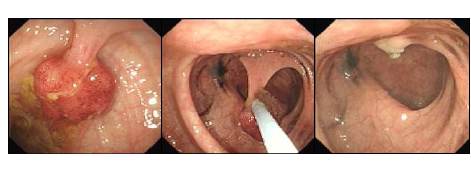
| 17:12, 23 January 2018 | Polypectomy.jpg (file) |  |
185 KB | 1 | |
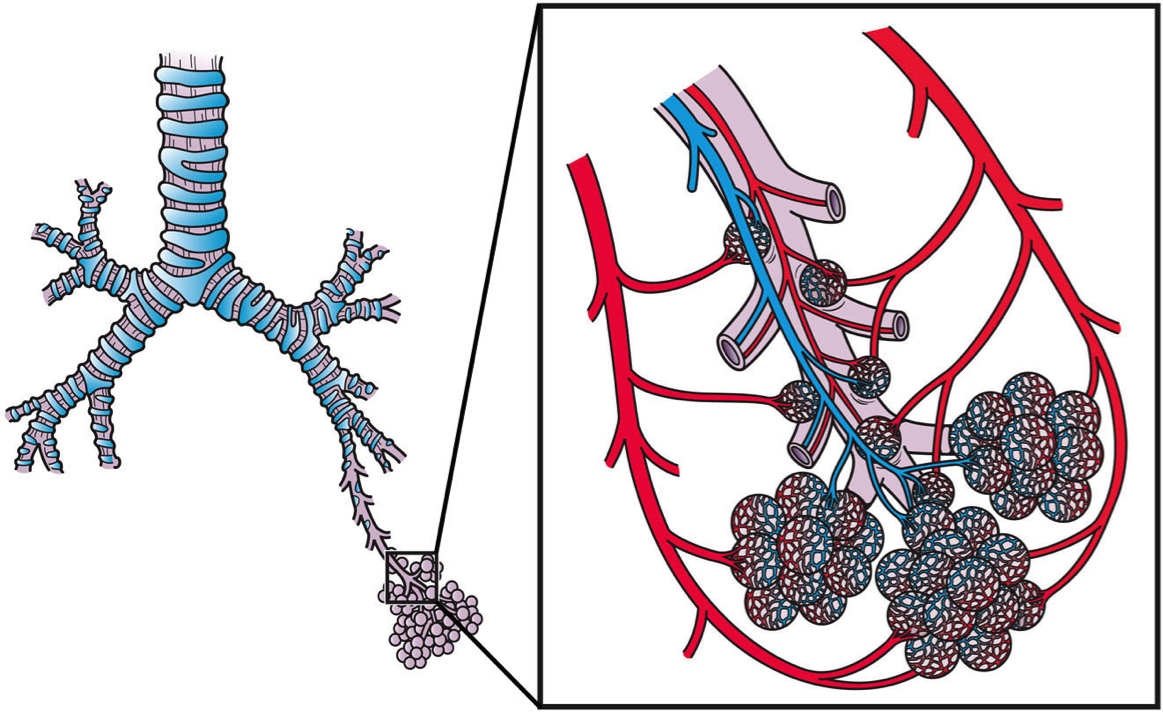
| 21:40, 12 February 2018 | Pulmonary Blood Circulation.png (file) |  |
776 KB | This slide shows the arterial and venous blood circulation of the pulmonary system. By Artwork by Holly Fischer - http://open.umich.edu/education/med/resources/second-look-series/materials - Respiratory Tract Slide 20, CC BY 3.0,<ref name="urlFile:Pulm... | 1 |
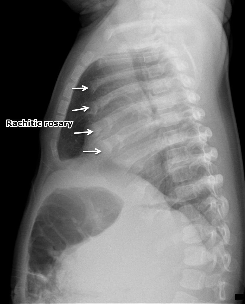
| 01:04, 26 November 2017 | Rachitic-rosary.png (file) |  |
529 KB | Rachitic rosary: Widening of the anterior rib ends at the costochondral junctions. Case courtesy of Dr Dalia Ibrahim, Radiopaedia.org, rID: 47584 https://radiopaedia.org/cases/47584 | 1 |
| 01:04, 26 November 2017 | Rachitic-rosary (1).png (file) | .png) |
433 KB | Rachitic rosary: Widening of the anterior rib ends at the costochondral junctions. Case courtesy of Dr Dalia Ibrahim, Radiopaedia.org, rID: 47584 https://radiopaedia.org/cases/47584 | 1 |
| 15:19, 4 December 2017 | ResectedIleum.jpg (file) |  |
137 KB | Terminal ileum resected for Crohn's disease. By PPSE15 - Own work, CC BY-SA 4.0<ref name="urlFile:ResectedIleum.jpg - Wikimedia Commons">{{cite web |url=https://commons.wikimedia.org/w/index.php?curid=39360128 |title=File:ResectedIleum.jpg - Wikimedia... | 1 |
| 23:39, 25 November 2017 | Rickets-16.png (file) |  |
521 KB | Rickets: Metaphyseal flaring and fraying seen at both proximal and distal tibia. There is slight outward bowing of both tibias. Case courtesy of Dr Henry Knipe, Radiopaedia.org, rID: 41684 https://radiopaedia.org/cases/41684 | 1 |
| 23:57, 25 November 2017 | Rickets-2.png (file) |  |
829 KB | Case courtesy of A.Prof Frank Gaillard, Radiopaedia.org, rID: 8225 https://radiopaedia.org/cases/8225 The physes are widened with metaphyseal flaring. Note how the bones are coarse. | 1 |
| 23:51, 25 November 2017 | Rickets-with-pathological-fracture.png (file) |  |
204 KB | Pathological fracture of the femur shaft. The fracture seems weeks old with beginning callus. however, it is only partially consolidated. The main impression is the saber-like appearance of the bone which is common for patients with vitamin D deficienc... | 1 |
| 01:57, 16 May 2017 | SadafSharfaei.jpg (file) |  |
906 KB | 1 | |
| 16:42, 31 January 2018 | Sagittal CT - FAP.jpg (file) |  |
73 KB | Sagittal CT shows multiple filling defects in a patient with familial adenomatous polyposis. Case courtesy of Dr David Cuete, Radiopaedia.org | 1 |
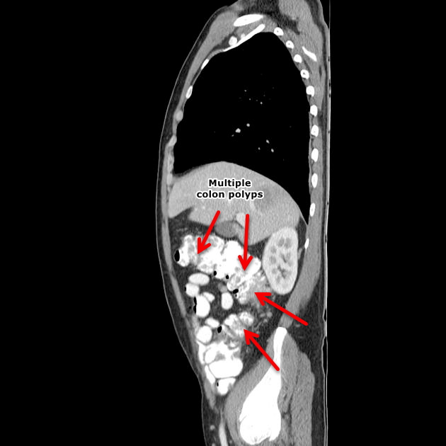
| 15:03, 31 January 2018 | Sagittal CT Colon polyps.jpeg (file) |  |
49 KB | Sagittal CT scan shows colon polyp | 1 |
| 19:29, 9 November 2017 | Small bilateral pleural effusions.jpg (file) |  |
146 KB | 1 | |
| 16:05, 27 October 2017 | Tropheryma whipplei.jpeg (file) |  |
52 KB | 1 | |
| 17:58, 23 January 2018 | Villous adenoma of the sigmoid colon, gross pathology.jpg (file) |  |
64 KB | 1 | |
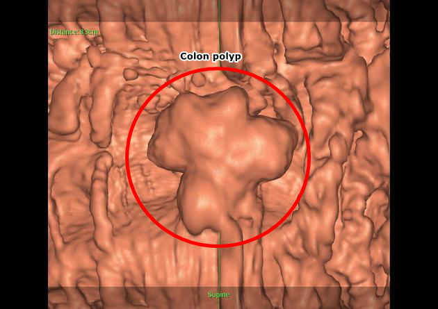
| 15:01, 31 January 2018 | Virtual colonoscopy Colon polyp.jpeg (file) |  |
116 KB | Virtual colonoscopy shows pedunculated colon polyp-Case courtesy of Dr Ayaz Hidayatov, Radiopaedia.org | 1 |
| 15:57, 17 November 2017 | Whipple's disease.gif (file) |  |
552 KB | 3 | |
| 16:03, 17 November 2017 | Whipple's disease1.gif (file) |  |
552 KB | 1 | |
| 19:42, 17 October 2017 | Whipple disease - intermed mag.jpg (file) |  |
273 KB | Intermediate magnification micrograph of Whipple's disease, also Whipple disease. H&E stain. Duodenal biopsy. The images show the characteristic feature of Whipple's disease; foamy macrophages are present in the lamina propria. | 1 |
| 15:05, 16 November 2017 | Whipple disease Acid fast stain.jpg (file) |  |
847 KB | 1 | |
| 14:59, 16 November 2017 | Whipple disease GMS stain.jpg (file) |  |
1.45 MB | 1 | |
| 15:01, 16 November 2017 | Whipple disease HE.jpg (file) |  |
1.96 MB | 1 | |
| 15:02, 16 November 2017 | Whipple disease PAS positive1.jpg (file) |  |
308 KB | 1 | |
| 15:02, 16 November 2017 | Whipple disease PAS positive2.jpg (file) |  |
252 KB | 1 | |
| 15:03, 16 November 2017 | Whipple disease PAS positive3.jpg (file) |  |
1.08 MB | 1 | |
| 14:58, 16 November 2017 | Whipple disease PAS stain-m1.jpg (file) |  |
1.49 MB | 1 | |
| 15:26, 15 November 2017 | Whipple disease PAS stain 1.jpg (file) |  |
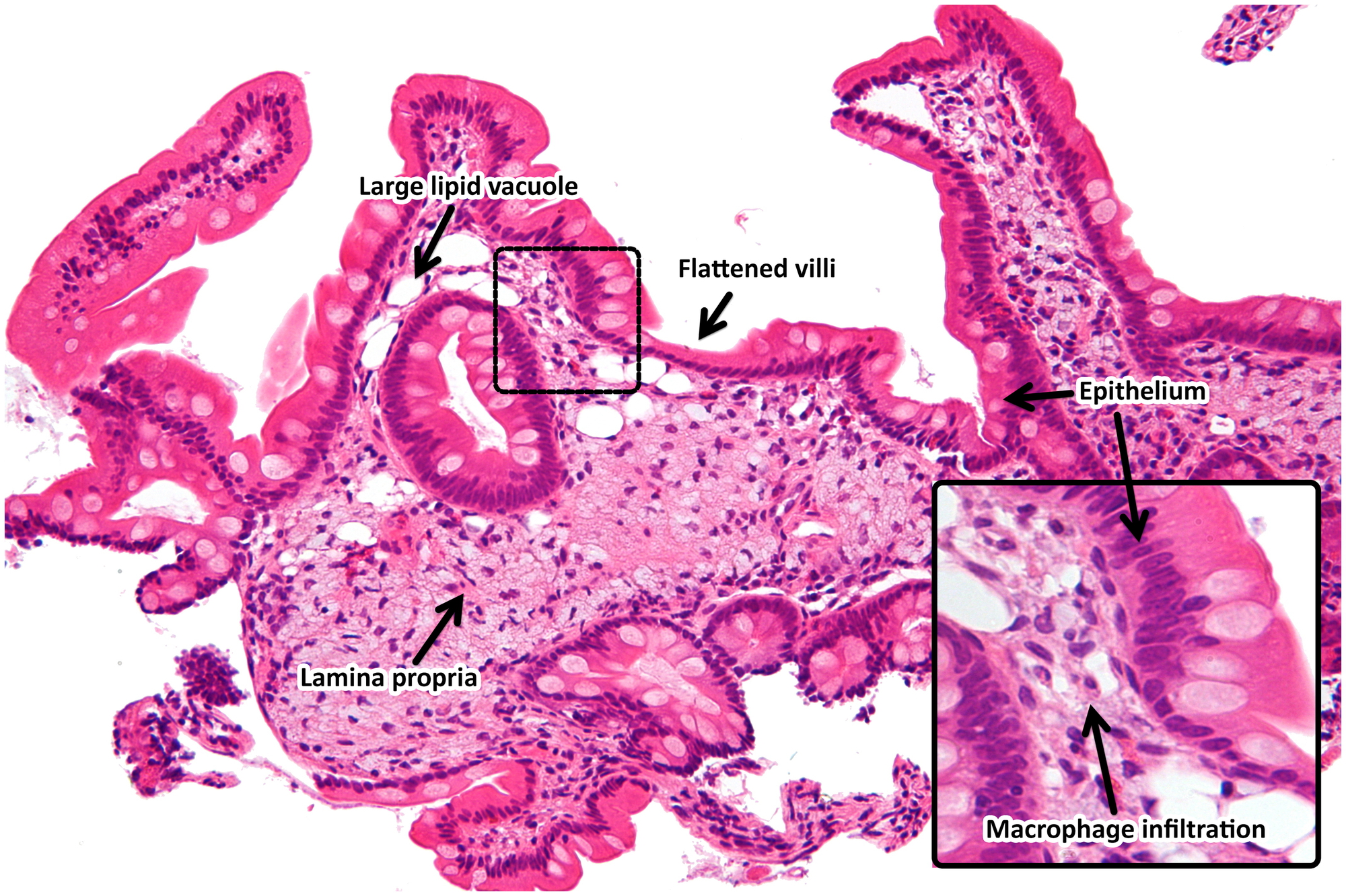
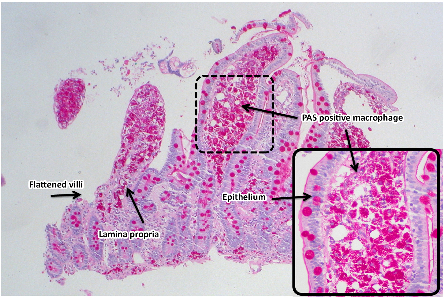
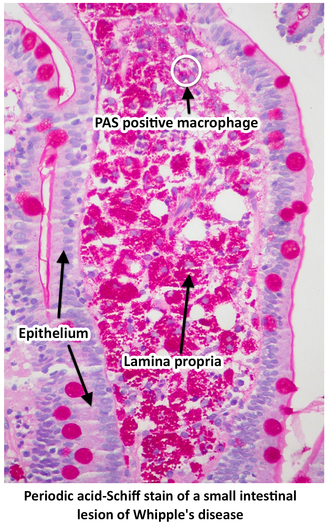
1.48 MB | Periodic acid-Schiff stain of a small intestinal lesion of Whipple's disease by Ed Uthman from Houston, TX, USA - Whipple's Disease, PAS https://commons.wikimedia.org/w/index.php?curid=30104677 | 1 |
| 15:32, 15 November 2017 | Whipple disease PAS stain 2.jpg (file) |  |
1.42 MB | 1 | |
| 15:32, 15 November 2017 | Whipple disease PAS stain 3.jpg (file) |  |
1.83 MB | 1 | |
| 19:43, 17 October 2017 | Whipple disease high mag.jpg (file) |  |
199 KB | High magnification micrograph of Whipple's disease, also Whipple disease. H&E stain. Duodenal biopsy. The images show the characteristic feature of Whipple's disease; foamy macrophages are present in the lamina propria. | 1 |
| 19:36, 17 October 2017 | Whipple disease low mag.jpg (file) |  |
1.52 MB | Low magnification micrograph of Whipple's disease. H&E stain. Duodenal biopsy. The images show the characteristic feature of Whipple's disease; foamy macrophages are present in the lamina propria. | 1 |
| 19:44, 17 October 2017 | Whipple disease very high mag.jpg (file) |  |
308 KB | Very high magnification micrograph of Whipple's disease, also Whipple disease. H&E stain. Duodenal biopsy. The images show the characteristic feature of Whipple's disease; foamy macrophages are present in the lamina propria. | 1 |