Uploads by Ssharfaei
Jump to navigation
Jump to search
This special page shows all uploaded files.
| Date | Name | Thumbnail | Size | Description | Versions |
|---|---|---|---|---|---|
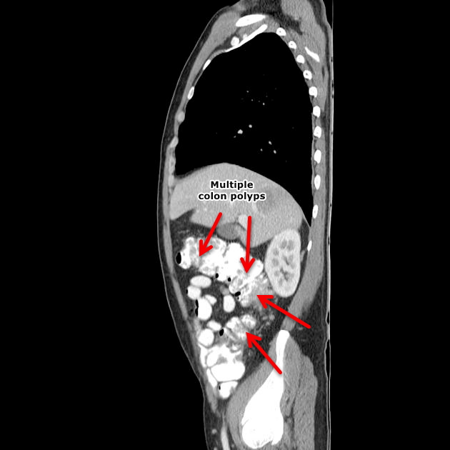
| 16:42, 31 January 2018 | Sagittal CT - FAP.jpg (file) |  |
73 KB | Sagittal CT shows multiple filling defects in a patient with familial adenomatous polyposis. Case courtesy of Dr David Cuete, Radiopaedia.org | 1 |
| 15:03, 31 January 2018 | Sagittal CT Colon polyps.jpeg (file) |  |
49 KB | Sagittal CT scan shows colon polyp | 1 |
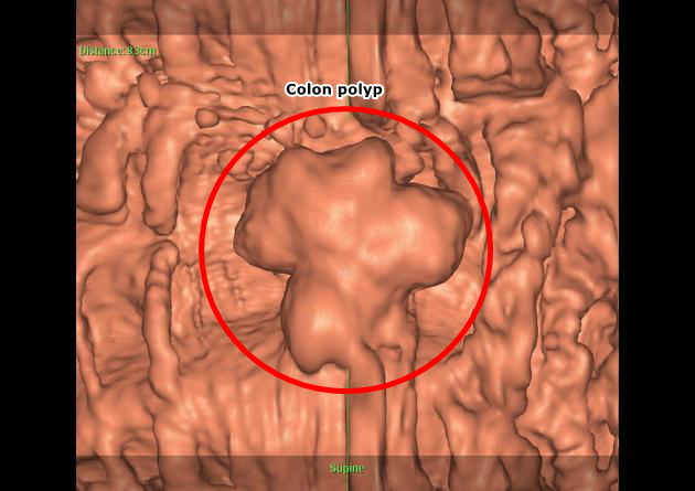
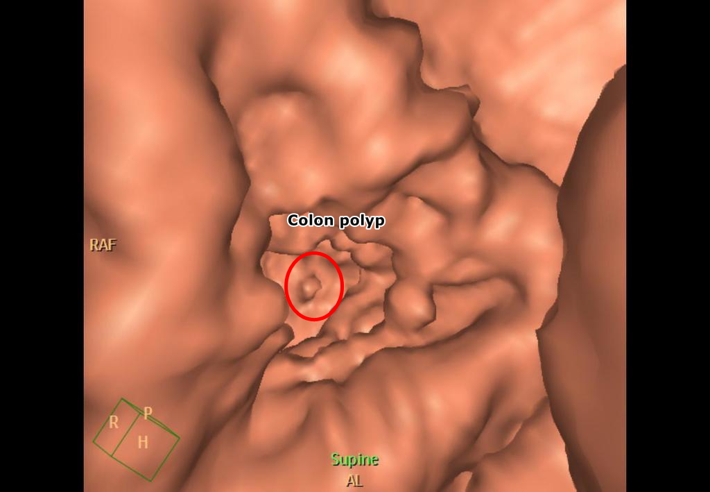
| 15:01, 31 January 2018 | Virtual colonoscopy Colon polyp.jpeg (file) |  |
116 KB | Virtual colonoscopy shows pedunculated colon polyp-Case courtesy of Dr Ayaz Hidayatov, Radiopaedia.org | 1 |
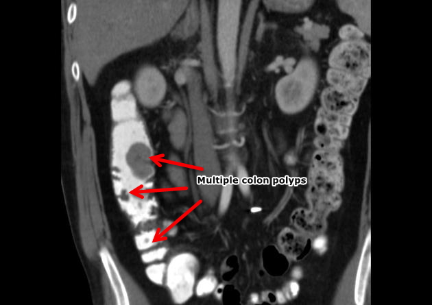
| 14:56, 31 January 2018 | Coronal CT colon polyps.jpeg (file) |  |
69 KB | Coronal CT scan shows multiple pedunculated colon polyps-Case courtesy of Dr Ayaz Hidayatov, Radiopaedia.org | 1 |
| 14:51, 31 January 2018 | 6fdbe648a73ff963226cfcf5c0414a big gallery.jpeg (file) |  |
76 KB | 1 | |
| 14:22, 31 January 2018 | Colonic-polyp-virtual-colonoscopy-1 (0).jpg (file) | .jpg) |
156 KB | 1 | |
| 15:59, 30 January 2018 | Familial adenomatous polyposis.jpg (file) |  |
78 KB | 1 | |
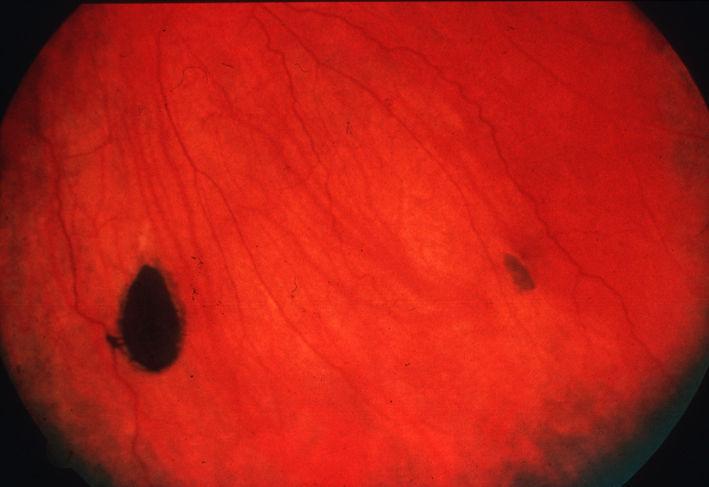
| 15:50, 30 January 2018 | CHRPE Congenital hypertrophy of the retinal pigment epithelium.jpg (file) |  |
31 KB | 1 | |
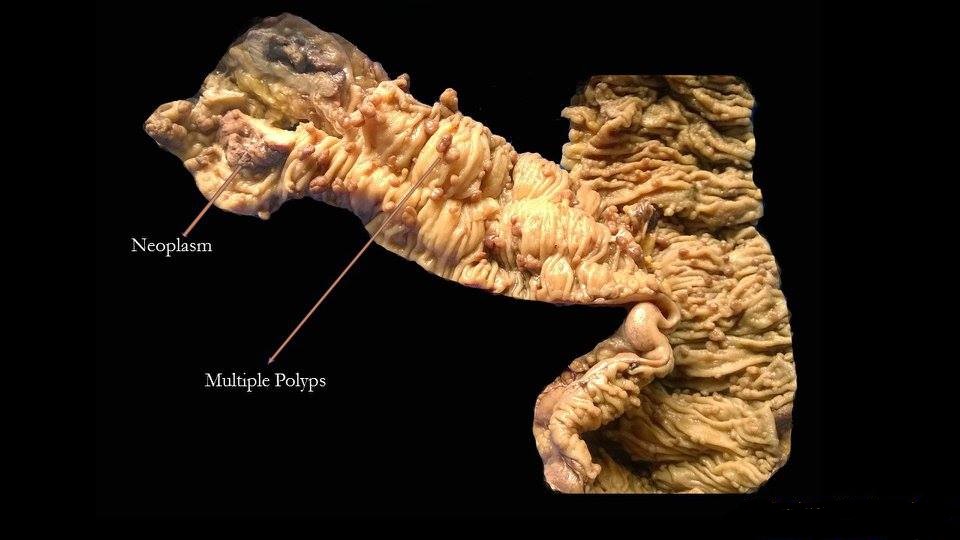
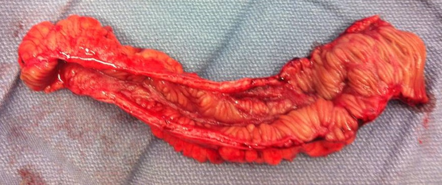
| 15:30, 30 January 2018 | Familial Adenomatous Polyposis intestine.jpg (file) |  |
497 KB | 2 | |
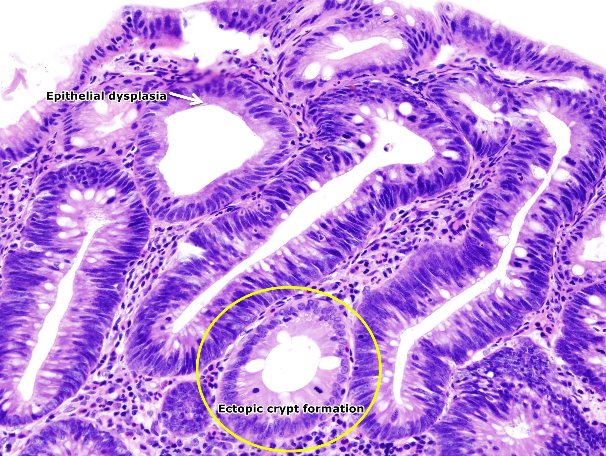
| 21:14, 26 January 2018 | Colon adenoma (1).jpg (file) | .jpg) |
1.81 MB | 3 | |
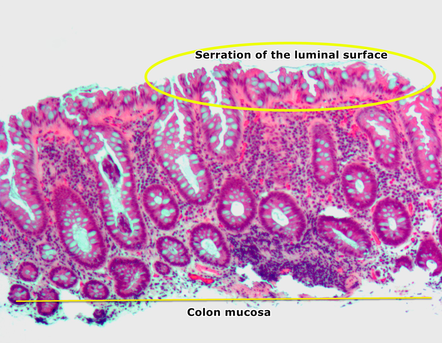
| 21:02, 26 January 2018 | Hyperplastic polyp2.jpg (file) |  |
946 KB | 3 | |
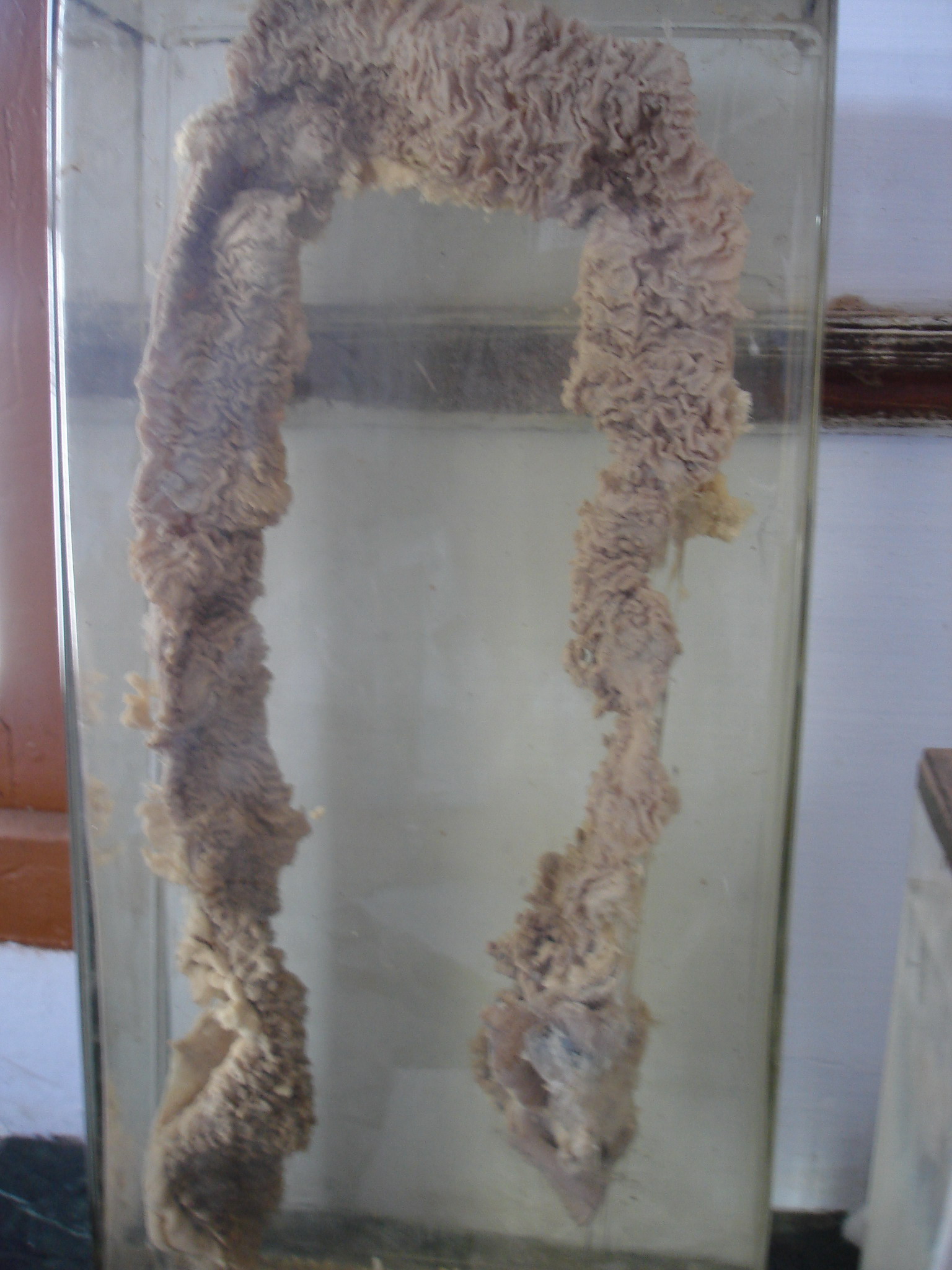
| 17:58, 23 January 2018 | Villous adenoma of the sigmoid colon, gross pathology.jpg (file) |  |
64 KB | 1 | |
| 17:43, 23 January 2018 | Colon-Polyp.jpg (file) |  |
778 KB | 1 | |
| 17:39, 23 January 2018 | Hyperplastic polyp of the colon, HE.png (file) |  |
1.93 MB | 1 | |
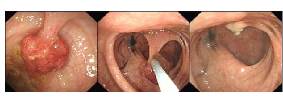
| 17:12, 23 January 2018 | Polypectomy.jpg (file) |  |
185 KB | 1 | |
| 17:12, 20 December 2017 | Image of resected colon segment with cancer & 4 nearby polyps plus schematic of field defects with sub-clones.jpg (file) |  |
625 KB | 1 | |
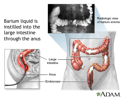
| 20:55, 19 December 2017 | Barium enema.jpg (file) |  |
81 KB | 1 | |
| 16:09, 5 December 2017 | Diagram of the small bowel 01 CRUK 045.jpg (file) |  |
254 KB | Diagram of the small bowel 01. Source: Wikimedia.org By Cancer Research UK - Original email from CRUK, CC BY-SA 4.0, https://commons.wikimedia.org/w/index.php?curid=34332940 | 1 |
| 14:32, 5 December 2017 | Crohn Jejunum.png (file) |  |
300 KB | Partial jejunum affected by morbus Crohn. Source: Wikimedia.org By Jaroslav Cehovsky - Camera, Public Domain, https://commons.wikimedia.org/w/index.php?curid=1458390 | 1 |
| 15:19, 4 December 2017 | ResectedIleum.jpg (file) |  |
137 KB | Terminal ileum resected for Crohn's disease. By PPSE15 - Own work, CC BY-SA 4.0<ref name="urlFile:ResectedIleum.jpg - Wikimedia Commons">{{cite web |url=https://commons.wikimedia.org/w/index.php?curid=39360128 |title=File:ResectedIleum.jpg - Wikimedia... | 1 |
| 01:04, 26 November 2017 | Rachitic-rosary (1).png (file) | .png) |
433 KB | Rachitic rosary: Widening of the anterior rib ends at the costochondral junctions. Case courtesy of Dr Dalia Ibrahim, Radiopaedia.org, rID: 47584 https://radiopaedia.org/cases/47584 | 1 |
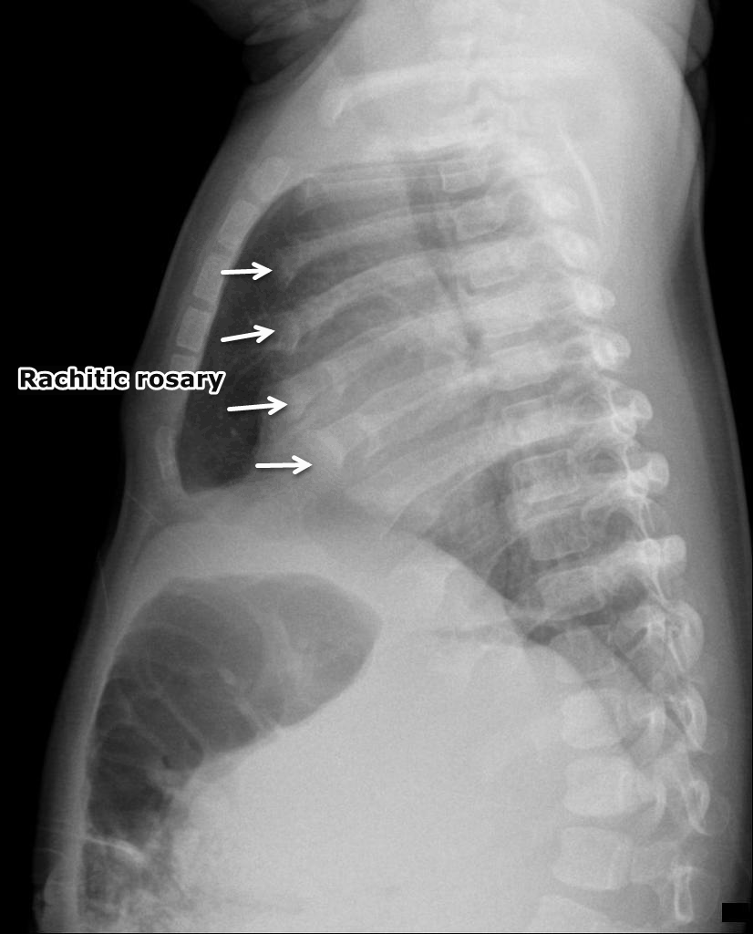
| 01:04, 26 November 2017 | Rachitic-rosary.png (file) |  |
529 KB | Rachitic rosary: Widening of the anterior rib ends at the costochondral junctions. Case courtesy of Dr Dalia Ibrahim, Radiopaedia.org, rID: 47584 https://radiopaedia.org/cases/47584 | 1 |
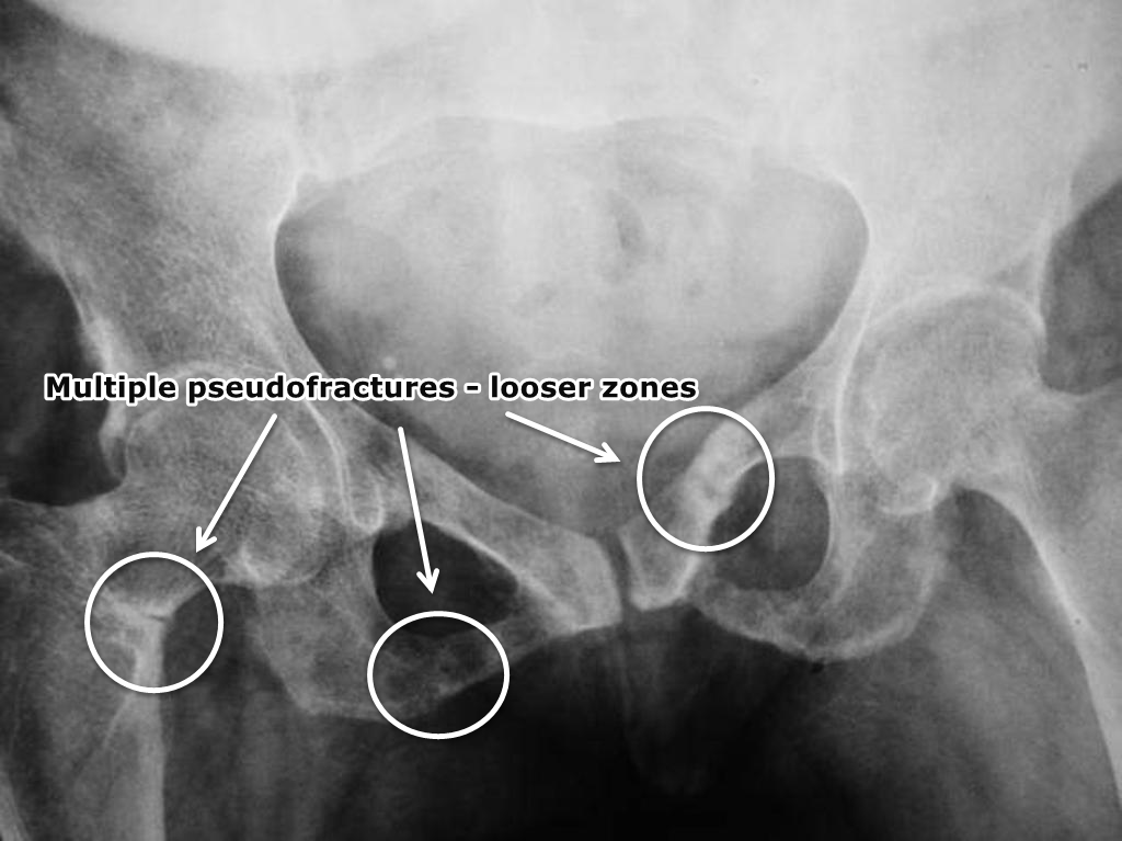
| 00:53, 26 November 2017 | Osteomalacia-looser-zones-1.png (file) |  |
452 KB | Osteopenic changes involving bony pelvis and proximal femurs. Multiple pseudofractures/Looser zones are seen involving superior and inferior pubic rami bilaterally. There is also a transcervical fracture on the right side. Case courtesy of Dr Iqbal Na... | 1 |
| 23:57, 25 November 2017 | Rickets-2.png (file) |  |
829 KB | Case courtesy of A.Prof Frank Gaillard, Radiopaedia.org, rID: 8225 https://radiopaedia.org/cases/8225 The physes are widened with metaphyseal flaring. Note how the bones are coarse. | 1 |
| 23:51, 25 November 2017 | Rickets-with-pathological-fracture.png (file) |  |
204 KB | Pathological fracture of the femur shaft. The fracture seems weeks old with beginning callus. however, it is only partially consolidated. The main impression is the saber-like appearance of the bone which is common for patients with vitamin D deficienc... | 1 |
| 23:39, 25 November 2017 | Rickets-16.png (file) |  |
521 KB | Rickets: Metaphyseal flaring and fraying seen at both proximal and distal tibia. There is slight outward bowing of both tibias. Case courtesy of Dr Henry Knipe, Radiopaedia.org, rID: 41684 https://radiopaedia.org/cases/41684 | 1 |
| 15:40, 22 November 2017 | LowKECG.png (file) |  |
1.24 MB | Hypokalemia | 1 |
| 21:37, 20 November 2017 | Anatomy-Male.ppt (file) | 285 KB | 2 | ||
| 21:33, 20 November 2017 | Anatomy-Female.ppt (file) | 770 KB | 2 | ||
| 16:03, 17 November 2017 | Whipple's disease1.gif (file) |  |
552 KB | 1 | |
| 15:57, 17 November 2017 | Whipple's disease.gif (file) |  |
552 KB | 3 | |
| 15:05, 16 November 2017 | Whipple disease Acid fast stain.jpg (file) |  |
847 KB | 1 | |
| 15:04, 16 November 2017 | Light microscopy of intestine-Whipples Disease.jpg .jpg (file) |  |
430 KB | 1 | |
| 15:03, 16 November 2017 | Whipple disease PAS positive3.jpg (file) |  |
1.08 MB | 1 | |
| 15:02, 16 November 2017 | Whipple disease PAS positive2.jpg (file) |  |
252 KB | 1 | |
| 15:02, 16 November 2017 | Whipple disease PAS positive1.jpg (file) |  |
308 KB | 1 | |
| 15:01, 16 November 2017 | Whipple disease HE.jpg (file) |  |
1.96 MB | 1 | |
| 14:59, 16 November 2017 | Whipple disease GMS stain.jpg (file) |  |
1.45 MB | 1 | |
| 14:58, 16 November 2017 | Whipple disease PAS stain-m1.jpg (file) |  |
1.49 MB | 1 | |
| 15:32, 15 November 2017 | Whipple disease PAS stain 3.jpg (file) |  |
1.83 MB | 1 | |
| 15:32, 15 November 2017 | Whipple disease PAS stain 2.jpg (file) |  |
1.42 MB | 1 | |
| 15:26, 15 November 2017 | Whipple disease PAS stain 1.jpg (file) |  |
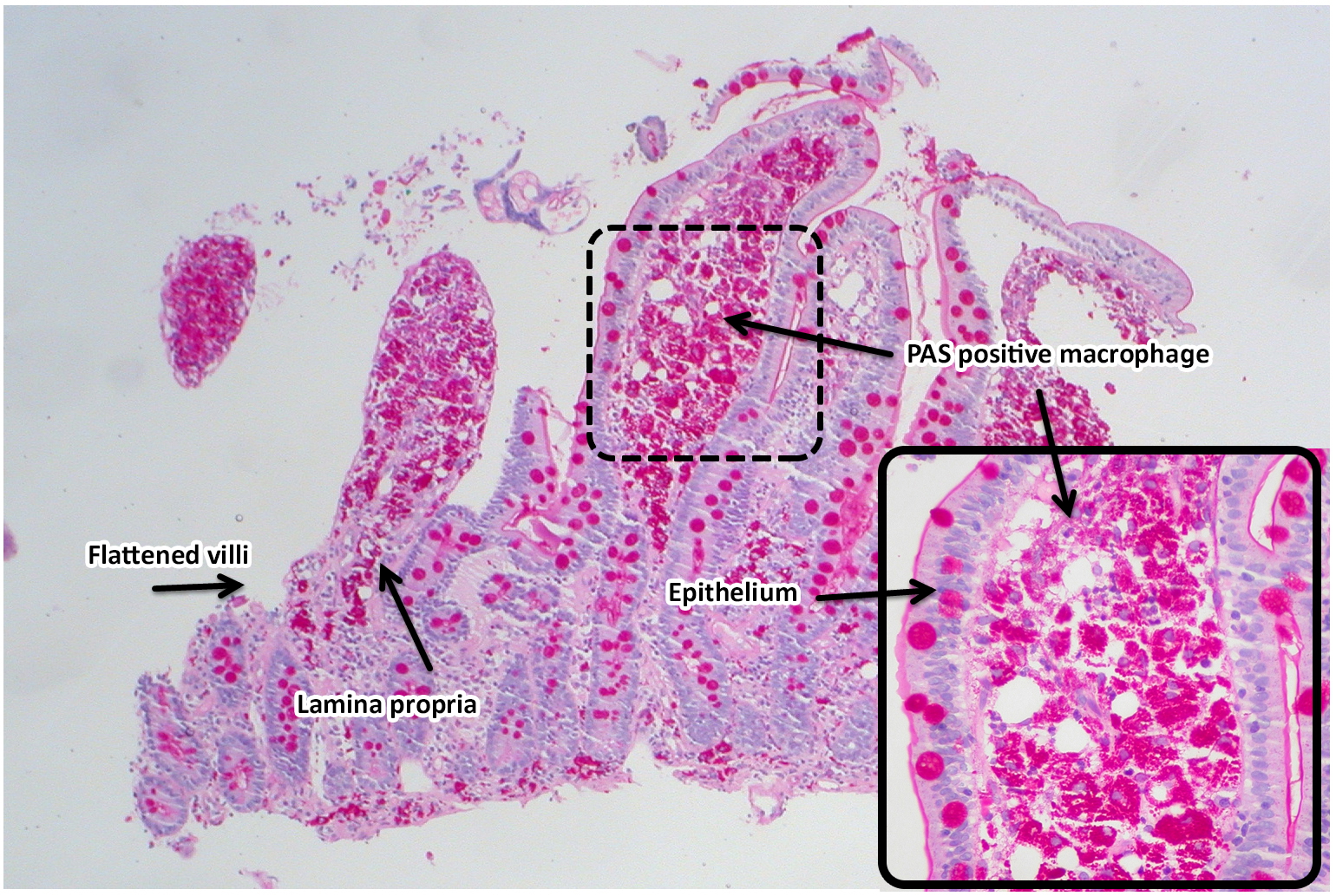
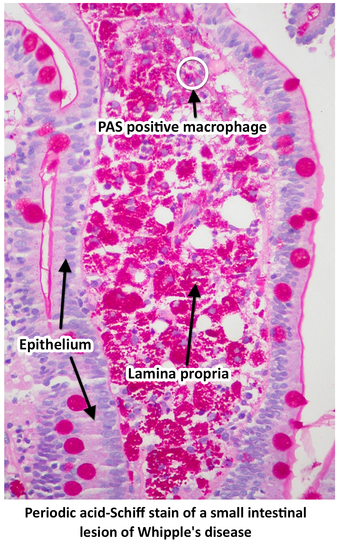
1.48 MB | Periodic acid-Schiff stain of a small intestinal lesion of Whipple's disease by Ed Uthman from Houston, TX, USA - Whipple's Disease, PAS https://commons.wikimedia.org/w/index.php?curid=30104677 | 1 |
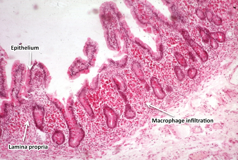
| 21:26, 13 November 2017 | Light microscopy of intestine-Whipples Disease.jpg (file) |  |
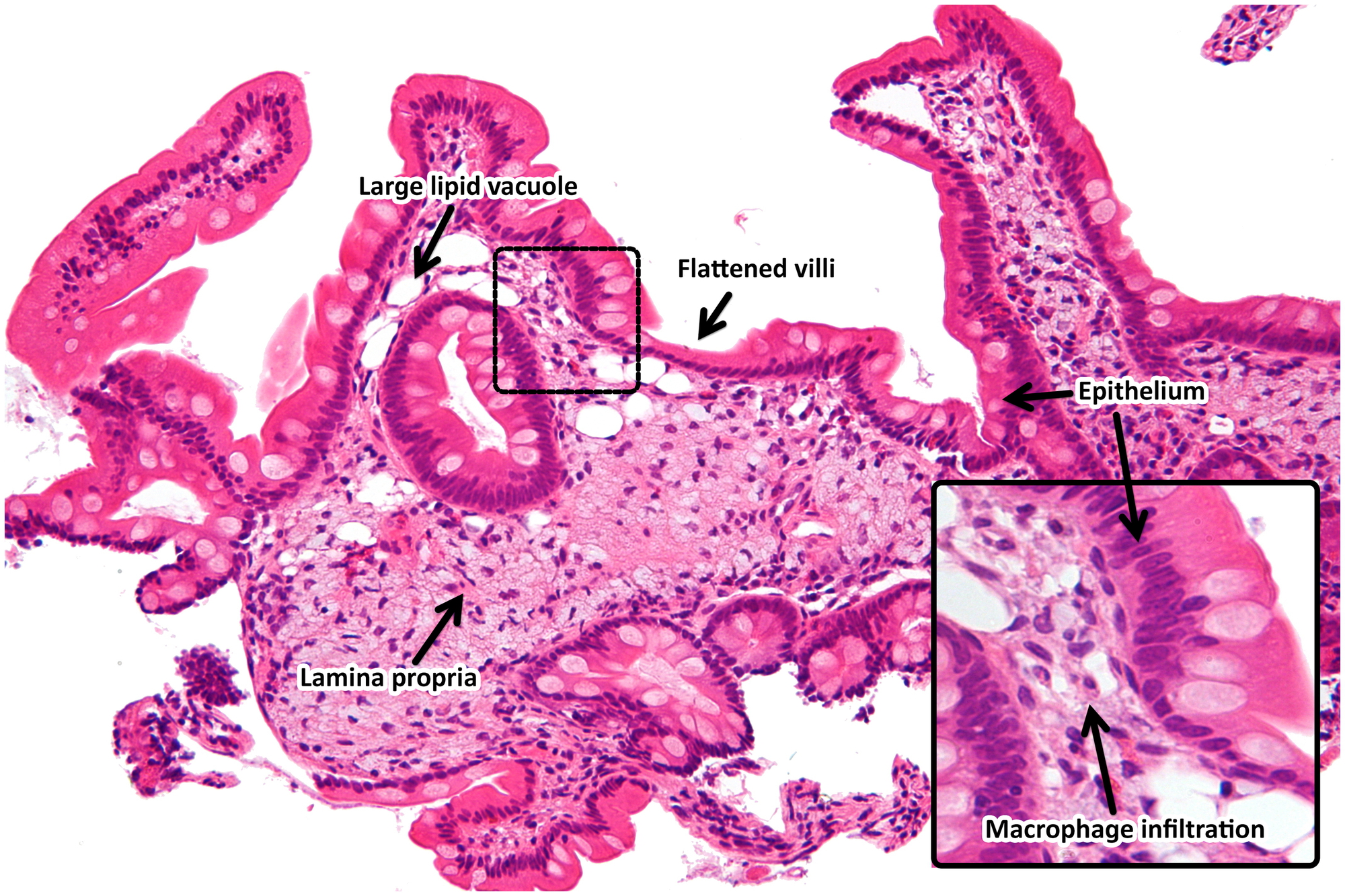
422 KB | light microscopy of intestine; Whipples Disease: Alcian blue with apparently eosin counterstain enlarged villus with many macrophages. From PEIR - University of Alabama at Birmingham Department of Pathology | 2 |
| 19:29, 9 November 2017 | Small bilateral pleural effusions.jpg (file) |  |
146 KB | 1 | |
| 16:05, 27 October 2017 | Tropheryma whipplei.jpeg (file) |  |
52 KB | 1 | |
| 19:44, 17 October 2017 | Whipple disease very high mag.jpg (file) |  |
308 KB | Very high magnification micrograph of Whipple's disease, also Whipple disease. H&E stain. Duodenal biopsy. The images show the characteristic feature of Whipple's disease; foamy macrophages are present in the lamina propria. | 1 |
| 19:43, 17 October 2017 | Whipple disease high mag.jpg (file) |  |
199 KB | High magnification micrograph of Whipple's disease, also Whipple disease. H&E stain. Duodenal biopsy. The images show the characteristic feature of Whipple's disease; foamy macrophages are present in the lamina propria. | 1 |
| 19:42, 17 October 2017 | Whipple disease - intermed mag.jpg (file) |  |
273 KB | Intermediate magnification micrograph of Whipple's disease, also Whipple disease. H&E stain. Duodenal biopsy. The images show the characteristic feature of Whipple's disease; foamy macrophages are present in the lamina propria. | 1 |
| 19:36, 17 October 2017 | Whipple disease low mag.jpg (file) |  |
1.52 MB | Low magnification micrograph of Whipple's disease. H&E stain. Duodenal biopsy. The images show the characteristic feature of Whipple's disease; foamy macrophages are present in the lamina propria. | 1 |
| 01:57, 16 May 2017 | SadafSharfaei.jpg (file) |  |
906 KB | 1 |