Second degree AV block EKG examples
Editor-In-Chief: C. Michael Gibson, M.S., M.D. [1] Syed Musadiq Ali M.B.B.S.[2]
For the main page on Second Degree AV Block, click here.
EKG Examples
Type I Second Degree AV Block
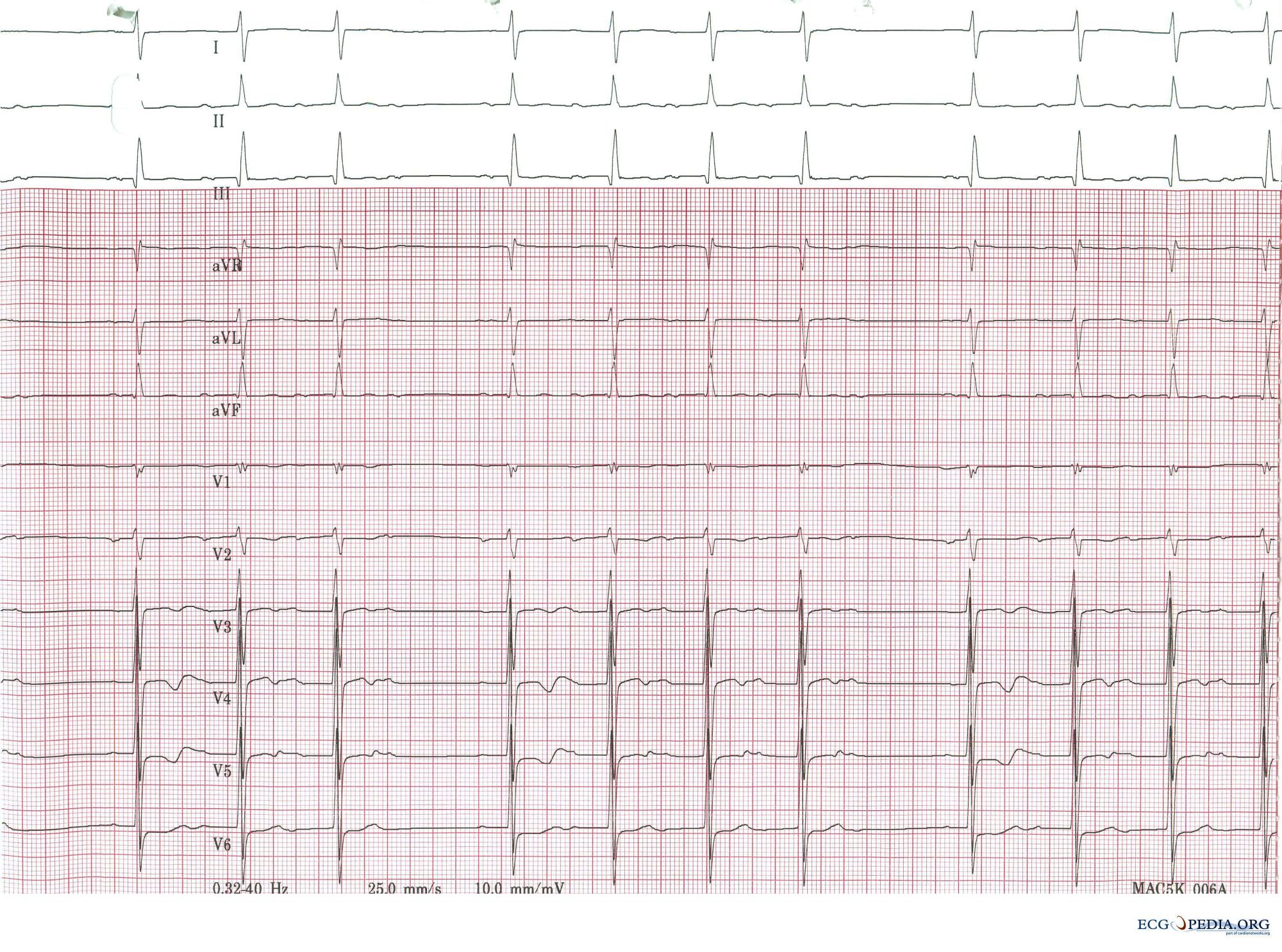
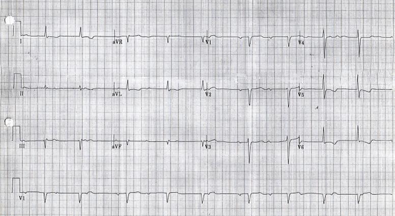
Shown below is an electrocardiogram showing type I second degree AV block (Wenckebach).

Copyleft image obtained courtesy of ECGpedia,http://en.ecgpedia.org/wiki/Main_Page
Shown below is an electrocardiogram showing type I second degree AV block (Wenckebach).

Copyleft image obtained courtesy of ECGpedia,http://en.ecgpedia.org/wiki/Main_Page
Shown below is an electrocardiogram showing type I second degree AV block (Wenckebach).

Copyleft image obtained courtesy of ECGpedia,http://en.ecgpedia.org/wiki/Main_Page
Shown below is an electrocardiogram showing a Mobitz I A/V block with a gradual increase in the PR interval before the dropped p wave

Copyleft image obtained courtesy of ECGpedia,http://en.ecgpedia.org/wiki/Main_Page
Type II Second-Degree AV Block (Mobitz Type II Block)
Shown below is an electrocardiogram of a 3 channel recording with a 2:1 AV block in a 73 year old woman with dizziness. 2 to 1 AV block (every other P wave is conducted to the ventricles) 2 to 1 AV block starts after the 5th QRS in this 3 channel recording. The first non-conducted P wave is indicated with an arrow. Note the long PR interval of conducted P waves is constant and the left bundle branch block 2 to 1 AV block cannot be classified into Mobitz type I or II as we do not know if the 2nd P wave would be conducted with the same or longer PR interval.

Copyleft image obtained courtesy of ECGpedia, http://en.ecgpedia.org/wiki/Main_Page
Shown below is an electrocardiogram of a 2:1 AV Block with atrial tachycardia.

Copyleft image obtained courtesy of ECGpedia, http://en.ecgpedia.org/wiki/Main_Page
Shown below is an example of EKG showing two strips from the same patient with a 2:1 block on the top tracing and a Mobitz II A/V block on the lower one. Note that with 2:1 block you cannot tell if this is a Mobitz I or II. Mobitz II is seen below as the PR does not change before and after the non-conducted P wave.

Copyleft image obtained courtesy of ECGpedia, http://en.ecgpedia.org/wiki/Main_Page