Sarcoidosis physical examination: Difference between revisions
No edit summary |
No edit summary |
||
| Line 1: | Line 1: | ||
__NOTOC__ | __NOTOC__ | ||
{{Sarcoidosis}} | {{Sarcoidosis}} | ||
{{CMG}}; {{AE}} | {{CMG}}; {{AE}}: Roshan Dinparasti Saleh M.D. | ||
==Overview== | ==Overview== | ||
Revision as of 16:20, 19 April 2018
|
Sarcoidosis Microchapters |
|
Diagnosis |
|---|
|
Treatment |
|
Case Studies |
|
Sarcoidosis physical examination On the Web |
|
American Roentgen Ray Society Images of Sarcoidosis physical examination |
|
Risk calculators and risk factors for Sarcoidosis physical examination |
Editor-In-Chief: C. Michael Gibson, M.S., M.D. [1]; Associate Editor(s)-in-Chief: : Roshan Dinparasti Saleh M.D.
Overview
Physical examination
Pulmonary Sarcoidosis
In half of the patients diagnosed with sarcoidosis, the disease is found incidentally by a CXR (bilateral hilar lymphadenopathy, reticular opacities) before the symptoms develop. Lung is the most common organ involved by sarcoidosis, but up to 30% percent of patients present with extra-pulmonary manifestations of sarcoidosis. The most common pattern of lung involvement in sarcoidosis is interstitial lung disease (other less common pulmonary manifestations include pneumothorax, pleural thickening, chylothorax, pulmonary hypertension)[1][2][3].
- Crackles are not commonly auscultated on lung examination. Wheezing may be heard when there is endobronchial involvement.
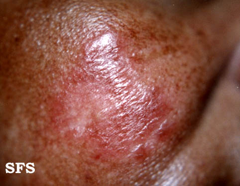
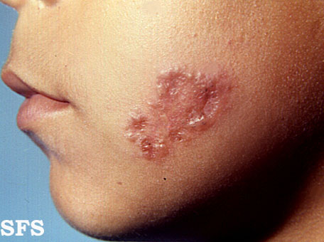
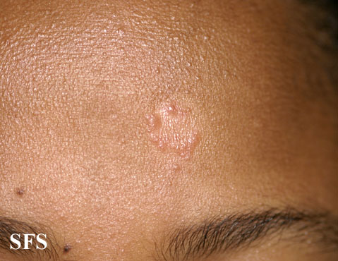
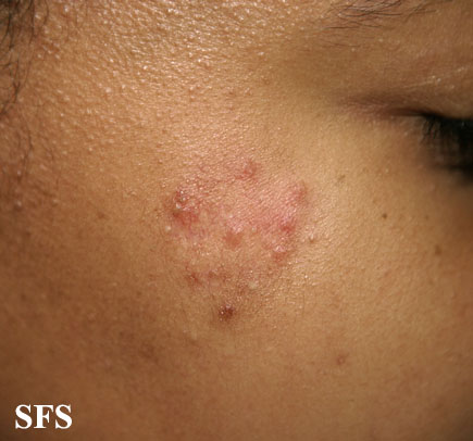
- In 8 to 15 year-old children, the disease presentation is similar to adults but younger children present with skin rash, arthritis, and red eye(uveitis). 90% of the children have an abnormal CXR[4][5][6].
Gallery
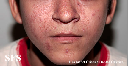
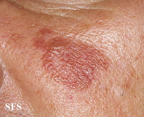
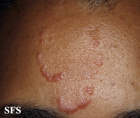
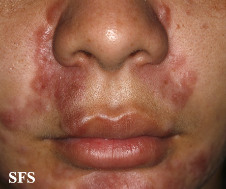
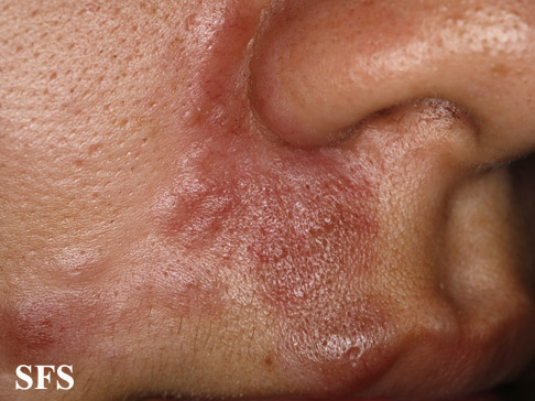
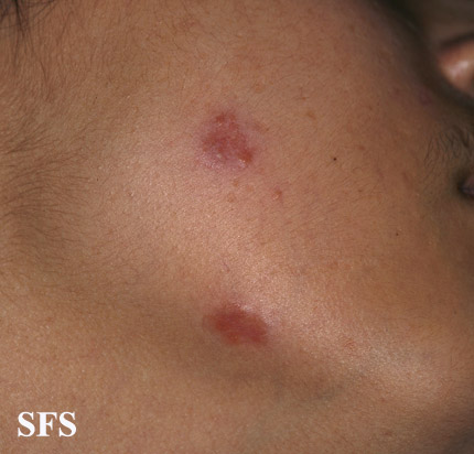
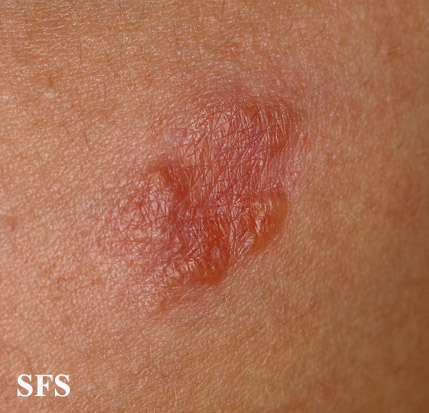
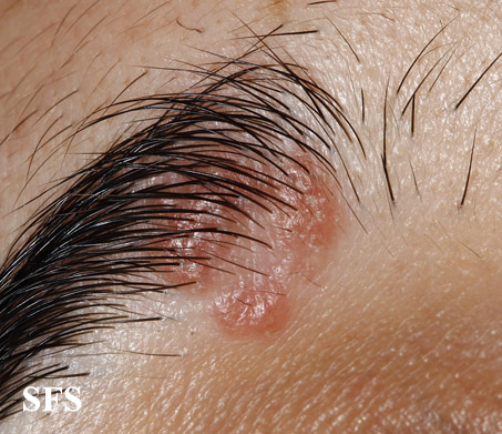
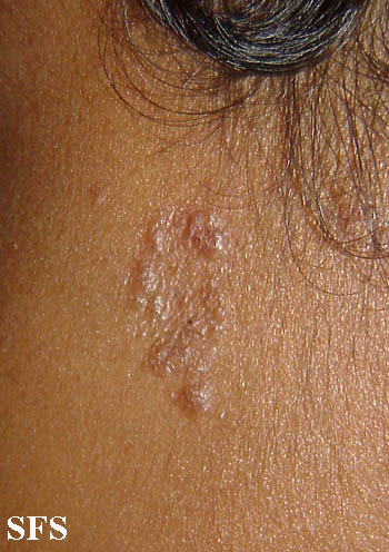
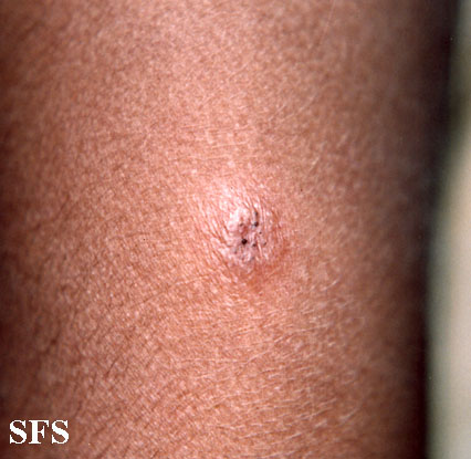
Skin
Head
Neck
Extremities
References
- ↑ Ungprasert P, Carmona EM, Utz JP, Ryu JH, Crowson CS, Matteson EL: Epidemiology of Sarcoidosis 1946-2013: A Population-Based Study. Mayo Clinic proceedings 2016, 91(2):183-188.
- ↑ Baughman RP, Teirstein AS, Judson MA, Rossman MD, Yeager H, Jr., Bresnitz EA, DePalo L, Hunninghake G, Iannuzzi MC, Johns CJ et al: Clinical characteristics of patients in a case control study of sarcoidosis. American journal of respiratory and critical care medicine 2001, 164(10 Pt 1):1885-1889.
- ↑ Rizzato G, Tinelli C: Unusual presentation of sarcoidosis. Respiration; international review of thoracic diseases 2005, 72(1):3-6.
- ↑ Nathan N, Marcelo P, Houdouin V, Epaud R, de Blic J, Valeyre D, Houzel A, Busson PF, Corvol H, Deschildre A et al: Lung sarcoidosis in children: update on disease expression and management. Thorax 2015, 70(6):537-542.
- ↑ Pattishall EN, Kendig EL, Jr.: Sarcoidosis in children. Pediatric pulmonology 1996, 22(3):195-203.
- ↑ Milman N, Hoffmann AL: Childhood sarcoidosis: long-term follow-up. The European respiratory journal 2008, 31(3):592-598.
