Sandbox:Javaria: Difference between revisions
No edit summary |
|||
| Line 1: | Line 1: | ||
__NOTOC__ | __NOTOC__ | ||
== | == Methods of Pacing == | ||
Cardiac pacemakers consist of two parts: a '''pulse generator''' or simply generator which is the source of electric pulse, and a variable number of '''leads''' that convey the electric signal from the generator to the [[Myocardium|myocardium]]. Newer leadless pacemakers have been introduced reducing the risk of complications. There is no formal classification system for the pacemaker. Based upon their use they can be divided into '''temporary pacing''' or emergency use pacing and '''permanent pacing'''. | |||
== | === Temporary pacing === | ||
Temporary pacing is indicated if permanent pacing is not instantly available, not required, or contraindicated. Different types of temporary pacing techniques include transvenous pacing, transcutaneous pacing, epicardial pacing, transesophageal atrial pacing<ref name="pmid16943920">{{cite journal |vauthors=Verbeet T, Castro J, Decoodt P |title=Transesophageal pacing: a versatile diagnostic and therapeutic tool |journal=Indian Pacing Electrophysiol J |volume=3 |issue=4 |pages=202–9 |date=October 2003 |pmid=16943920 |pmc=1502053 |doi= |url=}}</ref>, transthoracic mechanical pacing, transthoracic pacing<ref name="pmid6383140">{{cite journal |vauthors=Hedges JR, Syverud SA, Dalsey WC |title=Developments in transcutaneous and transthoracic pacing during bradyasystolic arrest |journal=Ann Emerg Med |volume=13 |issue=9 Pt 2 |pages=822–7 |date=September 1984 |pmid=6383140 |doi=10.1016/s0196-0644(84)80450-3 |url=}}</ref> and transmediastinal pacing. | |||
== | <br /> | ||
It | {| class="wikitable" | ||
! colspan=3 style="background: #4479BA; color: #FFFFFF; " align="center"|Types of temporary pacing | |||
|- | |||
!style="background: #4479BA; color: #FFFFFF; " align="center" |Type | |||
!style="background: #4479BA; color: #FFFFFF; " align="center" |Description | |||
!style="background: #4479BA; color: #FFFFFF; " align="center" |Indication | |||
|- | |||
|style="background: #DCDCDC; |Transvenous pacing {{main|Transvenous pacing}} | |||
|Transvenous pacing, when used for temporary pacing, is an alternative to transcutaneous pacing. A pacemaker wire is placed into a vein, under sterile conditions, and then passed into either the right atrium or right ventricle. The pacing wire is then connected to an external pacemaker outside the body. Transvenous pacing is often used as a bridge to permanent pacemaker placement. It can be kept in place until a permanent pacemaker is implanted or until there is no longer a need for a pacemaker and then it is removed. | |||
| | |||
|- | |||
|style="background: #DCDCDC; |Transcutaneous pacing{{main|Transcutaneous pacing}} | |||
|Transcutaneous pacing (TCP), also called external pacing, is recommended for the initial stabilization of hemodynamically significant [[bradycardia]]s of all types. The procedure is performed by placing two pacing pads on the patient's chest, either in the anterior/lateral position or the anterior/posterior position. The rescuer selects the pacing rate, and gradually increases the pacing current (measured in mA) until electrical capture (characterized by a wide QRS complex with a tall, broad T wave on the [[electrocardiogram|ECG]]) is achieved, with a corresponding pulse. Pacing artifact on the [[electrocardiogram|ECG]] and severe muscle twitching may make this determination difficult. External pacing should not be relied upon for an extended period of time. It is an emergency procedure that acts as a bridge until transvenous pacing or other therapies can be applied. | |||
| | |||
|- | |||
|style="background: #DCDCDC; |Epicardial Pacing{{Main|Epicardial}} | |||
|Temporary epicardial pacing is used during open heart surgery should the surgical procedure create [[Atrioventricular block|atrioventricular block]]. The electrodes are placed in contact with the [[Epicardium|outer wall]] of the ventricle to maintain satisfactory cardiac output until a temporary transvenous electrode has been inserted. | |||
| | |||
|- | |||
|style="background: #DCDCDC; |Transesophageal pacing | |||
| | |||
| | |||
|- | |||
|style="background: #DCDCDC; |Transthoracic pacing | |||
| | |||
| | |||
|- | |||
|style="background: #DCDCDC; |Transthoracic mechanical pacing | |||
|Also known as percussive pacing, is the use of the closed fist, usually on the left lower edge of the sternum over the right ventricle, striking from a distance of 20 - 30 cm to induce a ventricular beat (the British Journal of Anesthesia suggests this must be done to raise the ventricular pressure 10 - 15mmhg to induce electrical activity). This is an old procedure used only as a life saving means until an electrical pacemaker is brought to the patient.<ref>(Cite_Journal)Percussion pacing in a three year-old girl with complete heart block during cardiac catheterization. C Eich, A Bleckmann and T. Paul, retrieved from http://bja.oxfordjournals.org/cgi/content/full/95/4/465</ref> | |||
| | |||
|- | |||
|style="background: #DCDCDC; |Transmediastinal pacing | |||
| | |||
| | |||
|} | |||
==[[ | === Permanent Pacing === | ||
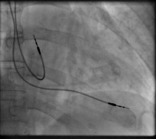
[[Image:Fluoroscopy pacemaker leads right atrium ventricle.png|thumb|left|Right atrial and right ventricular leads as visualized under X-ray during a pacemaker implant procedure. The atrial lead is the curved one making a U shape in the upper left part of the figure.]] | |||
Permanent pacing with an implantable pacemaker involves the transvenous placement of one or more pacing electrodes within a chamber, or chambers, of the heart. The procedure is performed by the incision of a suitable vein into which the electrode lead is inserted and passed along the vein, through the valve of the heart, until positioned in the chamber. The procedure is facilitated by [[fluoroscopy]] which enables the physician or cardiologist to view the passage of the electrode lead. After satisfactory lodgment of the electrode is confirmed the opposite end of the electrode lead is connected to the pacemaker generator. | |||
The pacemaker generator is a hermetically sealed device containing a power source, usually a lithium battery, a sensing amplifier which processes the electrical manifestation of naturally occurring heartbeats as sensed by the heart electrodes, the computer logic for the pacemaker and the output circuitry which delivers the pacing impulse to the electrodes. | |||
Most commonly, the generator is placed below the subcutaneous fat of the chest wall, above the muscles and bones of the chest. However, the placement may vary on a case by case basis. | |||
The outer casing of pacemakers is so designed that it will rarely be rejected by the body's [[immune system]]. It is usually made of [[titanium]], which is inert in the body. | |||
The | |||
Revision as of 13:03, 6 June 2020
Methods of Pacing
Cardiac pacemakers consist of two parts: a pulse generator or simply generator which is the source of electric pulse, and a variable number of leads that convey the electric signal from the generator to the myocardium. Newer leadless pacemakers have been introduced reducing the risk of complications. There is no formal classification system for the pacemaker. Based upon their use they can be divided into temporary pacing or emergency use pacing and permanent pacing.
Temporary pacing
Temporary pacing is indicated if permanent pacing is not instantly available, not required, or contraindicated. Different types of temporary pacing techniques include transvenous pacing, transcutaneous pacing, epicardial pacing, transesophageal atrial pacing[1], transthoracic mechanical pacing, transthoracic pacing[2] and transmediastinal pacing.
| Types of temporary pacing | ||
|---|---|---|
| Type | Description | Indication |
| Transvenous pacing | Transvenous pacing, when used for temporary pacing, is an alternative to transcutaneous pacing. A pacemaker wire is placed into a vein, under sterile conditions, and then passed into either the right atrium or right ventricle. The pacing wire is then connected to an external pacemaker outside the body. Transvenous pacing is often used as a bridge to permanent pacemaker placement. It can be kept in place until a permanent pacemaker is implanted or until there is no longer a need for a pacemaker and then it is removed. | |
| Transcutaneous pacing | Transcutaneous pacing (TCP), also called external pacing, is recommended for the initial stabilization of hemodynamically significant bradycardias of all types. The procedure is performed by placing two pacing pads on the patient's chest, either in the anterior/lateral position or the anterior/posterior position. The rescuer selects the pacing rate, and gradually increases the pacing current (measured in mA) until electrical capture (characterized by a wide QRS complex with a tall, broad T wave on the ECG) is achieved, with a corresponding pulse. Pacing artifact on the ECG and severe muscle twitching may make this determination difficult. External pacing should not be relied upon for an extended period of time. It is an emergency procedure that acts as a bridge until transvenous pacing or other therapies can be applied. | |
| Epicardial Pacing | Temporary epicardial pacing is used during open heart surgery should the surgical procedure create atrioventricular block. The electrodes are placed in contact with the outer wall of the ventricle to maintain satisfactory cardiac output until a temporary transvenous electrode has been inserted. | |
| Transesophageal pacing | ||
| Transthoracic pacing | ||
| Transthoracic mechanical pacing | Also known as percussive pacing, is the use of the closed fist, usually on the left lower edge of the sternum over the right ventricle, striking from a distance of 20 - 30 cm to induce a ventricular beat (the British Journal of Anesthesia suggests this must be done to raise the ventricular pressure 10 - 15mmhg to induce electrical activity). This is an old procedure used only as a life saving means until an electrical pacemaker is brought to the patient.[3] | |
| Transmediastinal pacing | ||
Permanent Pacing

Permanent pacing with an implantable pacemaker involves the transvenous placement of one or more pacing electrodes within a chamber, or chambers, of the heart. The procedure is performed by the incision of a suitable vein into which the electrode lead is inserted and passed along the vein, through the valve of the heart, until positioned in the chamber. The procedure is facilitated by fluoroscopy which enables the physician or cardiologist to view the passage of the electrode lead. After satisfactory lodgment of the electrode is confirmed the opposite end of the electrode lead is connected to the pacemaker generator.
The pacemaker generator is a hermetically sealed device containing a power source, usually a lithium battery, a sensing amplifier which processes the electrical manifestation of naturally occurring heartbeats as sensed by the heart electrodes, the computer logic for the pacemaker and the output circuitry which delivers the pacing impulse to the electrodes.
Most commonly, the generator is placed below the subcutaneous fat of the chest wall, above the muscles and bones of the chest. However, the placement may vary on a case by case basis.
The outer casing of pacemakers is so designed that it will rarely be rejected by the body's immune system. It is usually made of titanium, which is inert in the body.
- ↑ Verbeet T, Castro J, Decoodt P (October 2003). "Transesophageal pacing: a versatile diagnostic and therapeutic tool". Indian Pacing Electrophysiol J. 3 (4): 202–9. PMC 1502053. PMID 16943920.
- ↑ Hedges JR, Syverud SA, Dalsey WC (September 1984). "Developments in transcutaneous and transthoracic pacing during bradyasystolic arrest". Ann Emerg Med. 13 (9 Pt 2): 822–7. doi:10.1016/s0196-0644(84)80450-3. PMID 6383140.
- ↑ (Cite_Journal)Percussion pacing in a three year-old girl with complete heart block during cardiac catheterization. C Eich, A Bleckmann and T. Paul, retrieved from http://bja.oxfordjournals.org/cgi/content/full/95/4/465