Salter-Harris fracture: Difference between revisions
(Created page with "__NOTOC__ {{SI}} {{CMG}} ==Overview== A '''Salter–Harris fracture''' is a fracture that involves the epiphyseal plate or growth plate of a bone. It is a common inju...") |
m (Bot: Removing from Primary care) |
||
| (One intermediate revision by one other user not shown) | |||
| Line 1: | Line 1: | ||
[[File:Salter-Harris classification.jpg|alt=Courtesy of DrMars, <https://www.wikidoc.org>|thumb|Salter-Harris classification]] | |||
__NOTOC__ | __NOTOC__ | ||
{{SI}} | {{SI}} | ||
==Overview== | {{CMG}}; {{AE}}[[User:DrMars|Mohammadmain Rezazadehsaatlou[2]]]. | ||
==Overview<ref name="pmid27206505">{{cite journal |vauthors=Cepela DJ, Tartaglione JP, Dooley TP, Patel PN |title=Classifications In Brief: Salter-Harris Classification of Pediatric Physeal Fractures |journal=Clin. Orthop. Relat. Res. |volume=474 |issue=11 |pages=2531–2537 |date=November 2016 |pmid=27206505 |pmc=5052189 |doi=10.1007/s11999-016-4891-3 |url=}}</ref>== | |||
Injuries leading to the bone fracture affecting the epiphyseal plate, or physis, are important and common in orthopedic medicine and the cause diagnostic and treatment challenges for orthopaedic surgeons. The related incidence rate of these fracture among pedicatric population is 15-20%. | |||
== Historical Perspective <ref name="pmid9150026">{{cite journal |vauthors=Loder RT, Swinford AE, Kuhns LR |title=The use of helical computed tomographic scan to assess bony physeal bridges |journal=J Pediatr Orthop |volume=17 |issue=3 |pages=356–9 |date=1997 |pmid=9150026 |doi= |url=}}</ref><ref name="pmid19461377">{{cite journal |vauthors=Leary JT, Handling M, Talerico M, Yong L, Bowe JA |title=Physeal fractures of the distal tibia: predictive factors of premature physeal closure and growth arrest |journal=J Pediatr Orthop |volume=29 |issue=4 |pages=356–61 |date=June 2009 |pmid=19461377 |doi=10.1097/BPO.0b013e3181a6bfe8 |url=}}</ref><ref name="pmid16670543">{{cite journal |vauthors=Rohmiller MT, Gaynor TP, Pawelek J, Mubarak SJ |title=Salter-Harris I and II fractures of the distal tibia: does mechanism of injury relate to premature physeal closure? |journal=J Pediatr Orthop |volume=26 |issue=3 |pages=322–8 |date=2006 |pmid=16670543 |doi=10.1097/01.bpo.0000217714.80233.0b |url=}}</ref><ref name="pmid20563820">{{cite journal |vauthors=Lemburg SP, Lilienthal E, Heyer CM |title=Growth plate fractures of the distal tibia: is CT imaging necessary? |journal=Arch Orthop Trauma Surg |volume=130 |issue=11 |pages=1411–7 |date=November 2010 |pmid=20563820 |doi=10.1007/s00402-010-1140-1 |url=}}</ref>== | |||
In 1863, Foucher JT was the first person who described the injuries affecting the epiphyseal plate. | |||
In 1895, Poland J, classified the injuries affecting the epiphyseal plat into the four types. | |||
In 1936 , Aitken AP, defined the specific differences of different types of physes based on their differences in: structure, location, weightbearing status, and susceptibility to injury. | |||
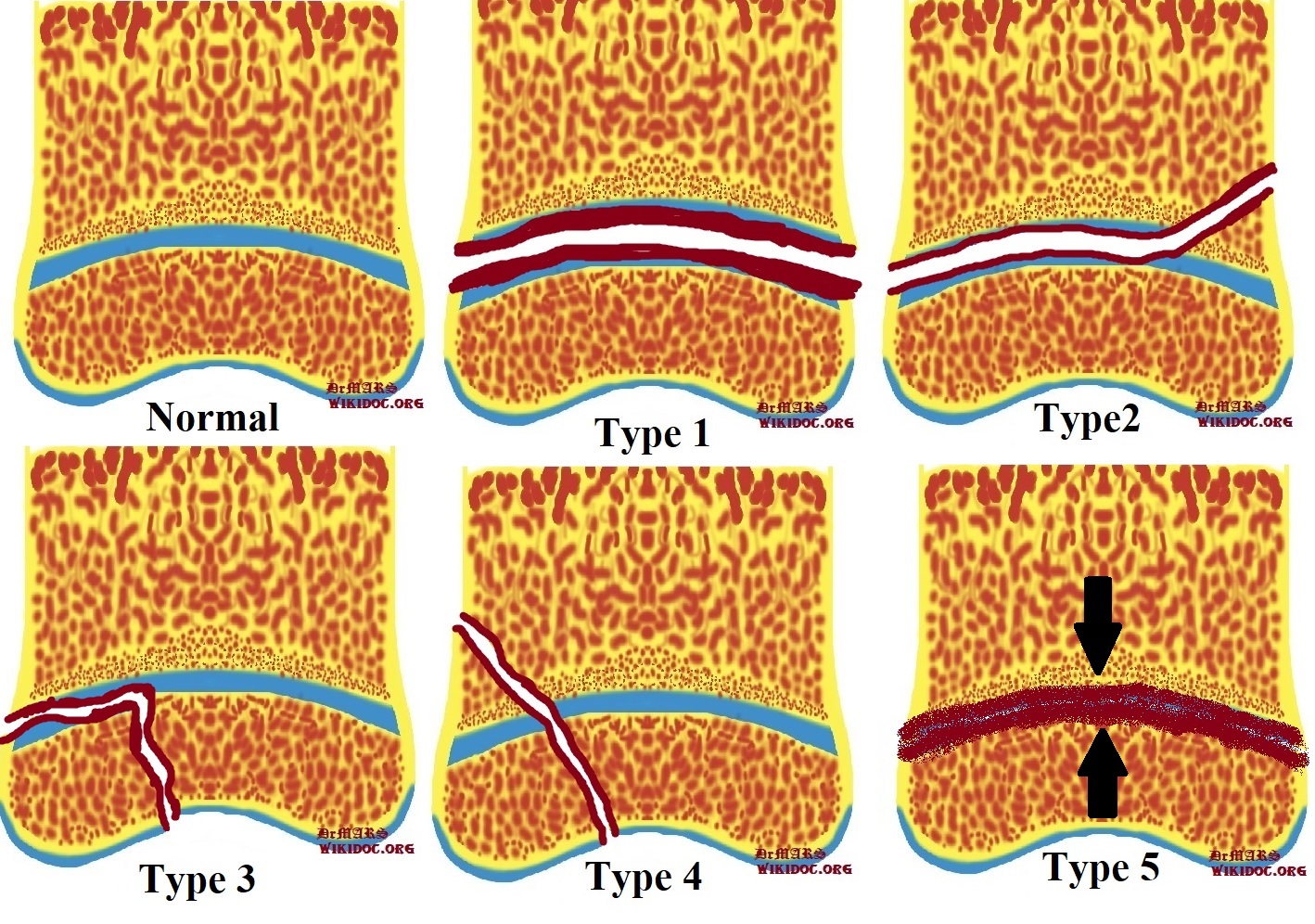
In 1963, two Canadian orthopaedic surgeons, Robert B. Salter (1924–2010) and W. Robert Harris (1922–2005), introduced a physeal fracture classification system according to the anatomy, fracture pattern, and prognosis of bone fracture. | |||
Then, various researchers and physicians tried to expanded the original work of Salter and Harris in order to make it to be to be more comprehensive: | |||
== | In 1968, Rang M, added a different sixth type of physeal injuries describing the caused damage to the perichondral ring due to the direct open injuries to the affected bone. | ||
{ | |||
In 1981, Ogden JA, described nine types of injuries such as injuries affecting the developing bone’s other growth mechanisms. | |||
==Salter-Harris classification== | |||
{| class="wikitable" | |||
|+ | |||
!Type | |||
!Descrpstion | |||
!Image | |||
!Radiography | |||
|- | |||
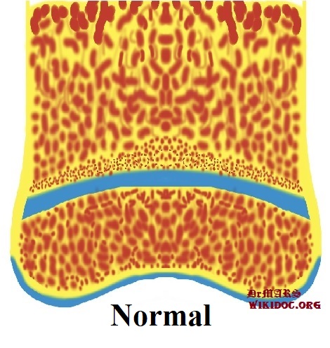
|Normal | |||
| | |||
|[[File:Normal Bone.jpg|alt=courtesy of DrMars, <https://www.wikidoc.org>|thumb|'''''Normal Bone''''']] | |||
| | |||
|- | |||
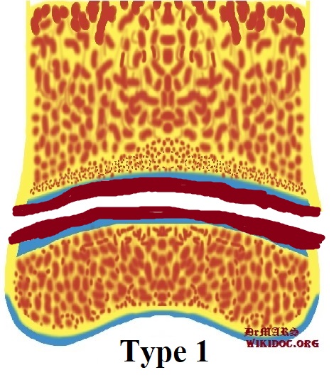
|Type I | |||
| | |||
* Frequency: 5-7% | |||
* cannot occur if the growth plate is fused cit | |||
* good prognosis | |||
* Mechanism: Fractured plane involved the whole growth plate, not involving bone | |||
* Origin: through the growth plate | |||
|[[File:Type1-Salter-Harris classification.jpg|alt=courtesy of DrMars, <https://www.wikidoc.org>|thumb|'''''Type 1-Salter-Harris classification''''']] | |||
|[[File:Salter-Harris type I injury of shoulder.jpg|thumb|'''Salter-Harris type I injury of shoulder''']] | |||
|- | |||
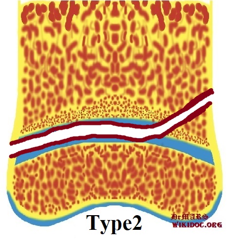
|Type II | |||
| | |||
* Frequency: 75% | |||
* good prognosis | |||
* Mechanism: Fractured plane involved most of the growth plate and up through the metaphysis | |||
* Origin: through the growth plate and the metaphysis, sparing the epiphysis | |||
|[[File:Type 2 Salter-Harris classification.jpg|alt=courtesy of DrMars, <https://www.wikidoc.org>|thumb|'''''Type 2 Salter-Harris classification''''']] | |||
|[[File:Salter-Harris type II injury of ankle.jpg|thumb|'''Salter-Harris type II injury of ankle''']] | |||
|- | |||
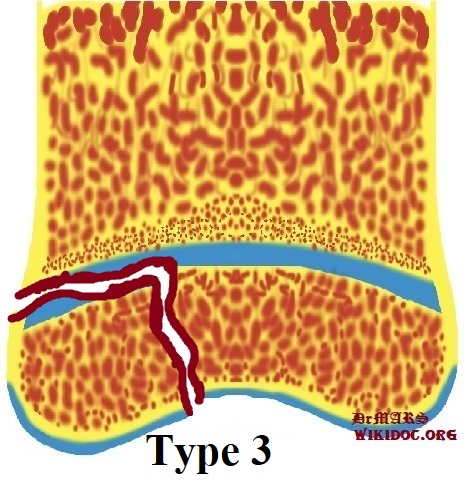
|Type III | |||
| | |||
* Frequency: 7-10% | |||
* cannot occur if the growth plate is fused cit | |||
* poorer prognosis as the proliferative and reserve zones are interrupted | |||
* Mechanism: Fractured plane involved the growth plate through the epiphysis | |||
* Origin: through growth plate and epiphysis, sparing the metaphysis | |||
|[[File:Type 3 Salter-Harris classification.jpg|alt=courtesy of DrMars, <https://www.wikidoc.org>|thumb|'''''Type 3 Salter-Harris classification''''']] | |||
|[[File:Salter-Harris type III injury of ankle.jpeg|thumb|'''Salter-Harris type III injury of ankle''']] | |||
|- | |||
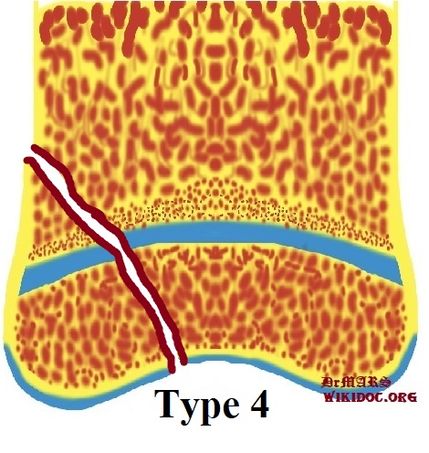
|Type IV | |||
| | |||
* Frequency: 10% | |||
* cannot occur intra-articular | |||
* poor prognosis as the proliferative and reserve zones are interrupted | |||
* Mechanism: Fractured plane involved directly through the metaphysis, growth plate and down through the epiphysis | |||
* Origin: through all three elements of the bone, the growth plate, metaphysis, and epiphysis | |||
|[[File:Type 4- Salter-Harris classification.jpg|alt=courtesy of DrMars, <https://www.wikidoc.org>|thumb|'''''Type 4- Salter-Harris classification''''']] | |||
|[[File:Salter-Harris type IV injury of foot.jpg|thumb|'''Salter-Harris type III injury of foot''']] | |||
|- | |||
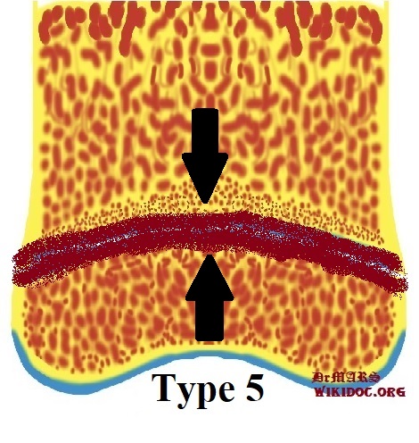
|Type V | |||
| | |||
* Frequency: <1% | |||
* cannot occur if the growth plate is fused cit | |||
* worst prognosis | |||
* Mechanism: Fractured plane dose note involved the growth plate but damages it by direct compression | |||
* Origin: decrease in the perceived space between the epiphysis and metaphysis | |||
|[[File:Type 5 Salter-Harris classification.jpg|alt=courtesy of DrMars, <https://www.wikidoc.org>|thumb|'''''Type 5 Salter-Harris classification''''']] | |||
| | |||
|- | |||
|Rare Types: Type VI | |||
| | |||
* An isolated damage of the perichondral structures | |||
| | |||
| | |||
|- | |||
|Rare Types: Type VII | |||
| | |||
* An isolated damage of the epiphyseal plate | |||
| | |||
| | |||
|- | |||
|Rare Types: Type VIII | |||
| | |||
* An isolated damage of the metaphysis, with a potential injury due to the endochondral ossification | |||
| | |||
| | |||
|- | |||
|Rare Types: Type IX | |||
| | |||
* An isolated damage of the periosteum that may interfere with membranous growth plane | |||
| | |||
| | |||
|} | |||
==See also== | |||
*[[Humerus fracture]] | |||
{{Fractures}} | {{Fractures}} | ||
[[ | |||
[[ | {{WikiDoc Sources}} | ||
[[ | |||
[[ | ==References== | ||
<references /> | |||
[[Category:Fractures]] | |||
[[Category:Injuries]] | |||
[[Category:Traumatology]] | |||
[[Category:Orthopedics]] | |||
[[Category:Disease]] | |||
[[Category:Radiology]] | |||
Latest revision as of 00:04, 30 July 2020
Editor-In-Chief: C. Michael Gibson, M.S., M.D. [1]; Associate Editor(s)-in-Chief: Mohammadmain Rezazadehsaatlou[2].
Overview[1]
Injuries leading to the bone fracture affecting the epiphyseal plate, or physis, are important and common in orthopedic medicine and the cause diagnostic and treatment challenges for orthopaedic surgeons. The related incidence rate of these fracture among pedicatric population is 15-20%.
Historical Perspective [2][3][4][5]
In 1863, Foucher JT was the first person who described the injuries affecting the epiphyseal plate.
In 1895, Poland J, classified the injuries affecting the epiphyseal plat into the four types.
In 1936 , Aitken AP, defined the specific differences of different types of physes based on their differences in: structure, location, weightbearing status, and susceptibility to injury.
In 1963, two Canadian orthopaedic surgeons, Robert B. Salter (1924–2010) and W. Robert Harris (1922–2005), introduced a physeal fracture classification system according to the anatomy, fracture pattern, and prognosis of bone fracture.
Then, various researchers and physicians tried to expanded the original work of Salter and Harris in order to make it to be to be more comprehensive:
In 1968, Rang M, added a different sixth type of physeal injuries describing the caused damage to the perichondral ring due to the direct open injuries to the affected bone.
In 1981, Ogden JA, described nine types of injuries such as injuries affecting the developing bone’s other growth mechanisms.
Salter-Harris classification
See also
References
- ↑ Cepela DJ, Tartaglione JP, Dooley TP, Patel PN (November 2016). "Classifications In Brief: Salter-Harris Classification of Pediatric Physeal Fractures". Clin. Orthop. Relat. Res. 474 (11): 2531–2537. doi:10.1007/s11999-016-4891-3. PMC 5052189. PMID 27206505.
- ↑ Loder RT, Swinford AE, Kuhns LR (1997). "The use of helical computed tomographic scan to assess bony physeal bridges". J Pediatr Orthop. 17 (3): 356–9. PMID 9150026.
- ↑ Leary JT, Handling M, Talerico M, Yong L, Bowe JA (June 2009). "Physeal fractures of the distal tibia: predictive factors of premature physeal closure and growth arrest". J Pediatr Orthop. 29 (4): 356–61. doi:10.1097/BPO.0b013e3181a6bfe8. PMID 19461377.
- ↑ Rohmiller MT, Gaynor TP, Pawelek J, Mubarak SJ (2006). "Salter-Harris I and II fractures of the distal tibia: does mechanism of injury relate to premature physeal closure?". J Pediatr Orthop. 26 (3): 322–8. doi:10.1097/01.bpo.0000217714.80233.0b. PMID 16670543.
- ↑ Lemburg SP, Lilienthal E, Heyer CM (November 2010). "Growth plate fractures of the distal tibia: is CT imaging necessary?". Arch Orthop Trauma Surg. 130 (11): 1411–7. doi:10.1007/s00402-010-1140-1. PMID 20563820.
