Rheumatoid arthritis x ray
|
Rheumatoid arthritis Microchapters | |
|
Diagnosis | |
|---|---|
|
Treatment | |
|
Case Studies | |
|
Rheumatoid arthritis x ray On the Web | |
|
American Roentgen Ray Society Images of Rheumatoid arthritis x ray | |
|
Risk calculators and risk factors for Rheumatoid arthritis x ray | |
Editor-In-Chief: C. Michael Gibson, M.S., M.D. [1] Associate Editor(s)-in-Chief: Manpreet Kaur, MD [2]
Overview
X Ray
Hallmark of rheumatoid arthritis are :
- Soft tissue swelling:
- This is an early finding in the course of rheumatoid arthritis.
- Soft tissue swelling is fusiform and periarticular results from of joint effusion, edema, and tenosynovitis.
- Joint space narrowing can be symmetrical or concentric.
- Marginal erosions can result from the erosion by pannus of the bone also called as “bare areas”
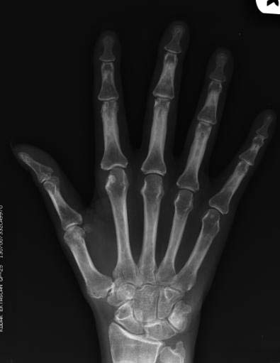
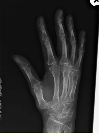
Hand and wrist findings
- Common joints involved are:
- Findings seen are:
Feet
- Various radiological findings are:
Shoulder
- Distal clavicle erosions
- Erosions of the superolateral aspect of the head of the humerus
- High riding shoulder due to subacromial-subdeltoid bursitis
Knee
- Joint effusions
- Loss of joint space
- Prepatellar bursitis
Hip
- Concentric loss of joint space
- Acetabular protrusio
Spine
- Atlantoaxial subluxation
- Atlantoaxial impaction: cephalad migration of C2
- Osteoporosis and osteoporotic fractures
- Erosion of spinous processes
-
-
-

-

-
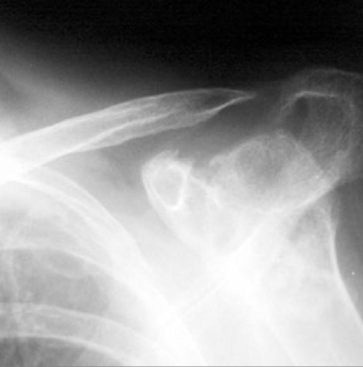
Distal clavicle erosion


