Rheumatoid arthritis x ray: Difference between revisions
(→X Ray) |
(→X Ray) |
||
| (30 intermediate revisions by 2 users not shown) | |||
| Line 4: | Line 4: | ||
==Overview== | ==Overview== | ||
The hallmark of [[rheumatoid arthritis]] is soft tissue swelling, joint space narrowing, and erosions. Hand and wrist findings on xray include subchondral [[cysts]], [[ulnar]] deviation of the [[MCP joint|MCP]] joints, [[boutonniere deformity|boutonniere]] and [[swan neck deformity|swan neck]] deformities, hitchhiker’s thumb deformity, scapholunate dissociation, ulnar translocation, and [[ankylosis]]. Feet findings on xray are [[subtalar joint]] involvement, posterior [[calcaneus|calcaneal]] tubercle erosion, hammer-toe deformity, and [[hallux]] [[valgus]]. Findings of shoulder such as distal [[clavicle]] erosions, erosions of the superolateral aspect of the head of the [[humerus]], and high riding [[shoulder]] due to subacromial-subdeltoid bursitis. Knee findings include [[joint]] effusions, loss of joint space, and [[prepatellar bursitis]]. Hip findings include concentric loss of [[joint]] space and acetabular protrusio. Spine findings are [[atlantoaxial]] subluxation, atlantoaxial impaction: cephalad migration of C2, [[osteoporosis]], and [[osteoporotic bones|osteoporotic]] fractures, and erosion of spinous processes. | |||
==X Ray== | ==X Ray== | ||
The hallmark of [[rheumatoid arthritis]] is:<ref name="pmid12477226">{{cite journal |vauthors=Devauchelle-Pensec V, Saraux A, Alapetite S, Colin D, Le Goff P |title=Diagnostic value of radiographs of the hands and feet in early rheumatoid arthritis |journal=Joint Bone Spine |volume=69 |issue=5 |pages=434–41 |date=October 2002 |pmid=12477226 |doi= |url=}}</ref> | |||
*Soft tissue swelling: | *Soft tissue swelling: | ||
** | **It is an early finding in the course of [[rheumatoid arthritis]]. | ||
**Soft tissue swelling is fusiform and periarticular | **Soft tissue swelling is [[fusiform]] and periarticular results from of joint effusion, [[edema]], and [[tenosynovitis]]. | ||
*Joint space narrowing can be symmetrical or concentric. | *[[Joint]] space narrowing: | ||
*Marginal erosions | **It can be symmetrical or [[concentric]]. | ||
'''Hand and wrist | *Marginal erosions: | ||
**These may result from the erosion by [[pannus]] of the [[bone]] also called as “bare areas”. | |||
*PIP and MCP joints (especially 2nd and 3rd MCP) | '''Hand and wrist''' | ||
*Ulnar styloid | |||
The common joints involved in a patient with rheumatoid arthritis, include: | |||
*PIP and [[MCP joint|MCP]] joints (especially 2nd and 3rd [[MCP joint|MCP]]) | |||
*[[Ulnar]] [[Styloid process|styloid]] | |||
*Triquetrum | *Triquetrum | ||
The findings may include:<ref name="pmid1731813">{{cite journal |vauthors=van der Heijde DM, van Leeuwen MA, van Riel PL, Koster AM, van 't Hof MA, van Rijswijk MH, van de Putte LB |title=Biannual radiographic assessments of hands and feet in a three-year prospective followup of patients with early rheumatoid arthritis |journal=Arthritis Rheum. |volume=35 |issue=1 |pages=26–34 |date=January 1992 |pmid=1731813 |doi= |url=}}</ref><ref name="pmid26247204">{{cite journal |vauthors=Koh JH, Jung SM, Lee JJ, Kang KY, Kwok SK, Park SH, Ju JH |title=Radiographic Structural Damage Is Worse in the Dominant than the Non-Dominant Hand in Individuals with Early Rheumatoid Arthritis |journal=PLoS ONE |volume=10 |issue=8 |pages=e0135409 |date=2015 |pmid=26247204 |pmc=4527732 |doi=10.1371/journal.pone.0135409 |url=}}</ref> | |||
*Subchondral cysts | *Subchondral [[cysts]] | ||
*Ulnar deviation of the MCP joints | *[[Ulnar]] deviation of the [[MCP joint|MCP]] joints | ||
*Boutonniere and swan neck deformities | *[[Boutonniere deformity|Boutonniere]] and [[Swan neck deformity|swan neck]] deformities | ||
*Hitchhiker’s thumb deformity | *Hitchhiker’s thumb deformity | ||
*Scapholunate dissociation, ulnar translocation | *Scapholunate dissociation, ulnar translocation | ||
*Ankylosis | *[[Ankylosis]] | ||
[[File:Rheumatoid-hands.jpg|200px|thumb|centre|Swan neck deformity <br>Source: A.Prof Frank Gaillard <ref> "https://radiopaedia.org, ref="https://radiopaedia.org/cases/7245">rID: 7245</ref>]] | |||
[[File:Rheumatoid-arthritis.jpg|200px|thumb|centre|Boutonniere deformity<br> Source:Case courtesy of Dr Hani Salam<ref> "https://radiopaedia.org/"Radiopaedia.org</ref>]] | |||
'''Feet''' | '''Feet''' | ||
Various radiological findings | |||
*Subtalar joint involvement | Various radiological findings of the feet that may be seen in rheumatoid arthritis include: | ||
*Posterior calcaneal tubercle erosion | *[[Subtalar joint|Subtalar]] joint involvement | ||
*Posterior [[Calcaneus|calcaneal]] tubercle erosion | |||
*Hammertoe deformity | *Hammertoe deformity | ||
*Hallux valgus | *[[Hallux]] [[valgus]] | ||
Image:Rheumatoid-arthritis- | [[Image:Rheumatoid-arthritis-feet.jpg|200px|centre|thumbnail|Marginal erosions involving the MTP joint of the right little toe<br> Source: Case courtesy of Dr Ian Bickle, <ref> href="https://radiopaedia.org/">Radiopaedia.org</ref><ref> href="https://radiopaedia.org/cases/27301">rID: 27301</ref>]] | ||
Image:Rheumatoid-arthritis- | |||
Image:Rheumatoid-arthritis- | '''Shoulder''' | ||
</ | Various radiological findings of the shoulder joint that may be seen in rheumatoid arthritis include: | ||
*Distal [[clavicle]] erosions | |||
*Erosions of the superolateral aspect of the head of the [[humerus]] | |||
*High riding [[shoulder]] due to subacromial-subdeltoid bursitis | |||
[[Image:Rheumatoid-arthritis-distal-clavicle-resorption.jpg|200px|centre|thumbnail|Distal clavicle resorption <br> Source:Case courtesy of Radswiki <ref>href="https://radiopaedia.org/">Radiopaedia.org</ref><ref> href="https://radiopaedia.org/cases/11884">rID: 11884</ref>]] | |||
'''Knee''' | |||
Various radiological findings of the knee joint that may be seen in rheumatoid arthritis include:<ref name="pmid2754663">{{cite journal |vauthors=Fuchs HA, Kaye JJ, Callahan LF, Nance EP, Pincus T |title=Evidence of significant radiographic damage in rheumatoid arthritis within the first 2 years of disease |journal=J. Rheumatol. |volume=16 |issue=5 |pages=585–91 |date=May 1989 |pmid=2754663 |doi= |url=}}</ref> | |||
*[[Joint]] effusions | |||
*Loss of joint space | |||
*[[Prepatellar bursitis]] | |||
'''Hip''' | |||
Various radiological findings of the hip joint that may be seen in rheumatoid arthritis include: | |||
*Concentric loss of [[joint]] space | |||
*Acetabular protrusio | |||
[[Image:Rheumatoid-arthritis-of-hip-with-erosion-and-pseudoarthrosis.jpg|centre|200px|thumbnail|Severe degenerative changes to the left hip with marked erosive change <br>Source:Case courtesy of Dr Jeremy Jones<ref> href="https://radiopaedia.org/">Radiopaedia.org</ref><ref> href="https://radiopaedia.org/cases/6464">rID: 6464</ref>]] | |||
'''Spinal cord''' | |||
Various radiological findings of the spinal cord joint that may be seen in rheumatoid arthritis include: | |||
*[[Atlantoaxial]] subluxation | |||
*Atlantoaxial impaction: cephalad migration of C2 | |||
*[[Osteoporosis]] and [[Osteoporotic bones|osteoporotic]] fractures | |||
*Erosion of spinous processes | |||
[[Image:Rheumatoid-arthritis-of-the-cervical-spine-1.jpg|200px|centre|thumbnail|Cervical spine<br> Source:Case courtesy of Dr Muhammad Essam, <ref> href="https://radiopaedia.org/">Radiopaedia.org</ref><ref> href="https://radiopaedia.org/cases/29388">rID: 29388</ref>]] | |||
==References== | ==References== | ||
[[Category:Needs content]] | [[Category:Needs content]] | ||
| Line 53: | Line 82: | ||
{{WH}} | {{WH}} | ||
<references /> | |||
Latest revision as of 17:24, 25 April 2018
|
Rheumatoid arthritis Microchapters | |
|
Diagnosis | |
|---|---|
|
Treatment | |
|
Case Studies | |
|
Rheumatoid arthritis x ray On the Web | |
|
American Roentgen Ray Society Images of Rheumatoid arthritis x ray | |
|
Risk calculators and risk factors for Rheumatoid arthritis x ray | |
Editor-In-Chief: C. Michael Gibson, M.S., M.D. [1] Associate Editor(s)-in-Chief: Manpreet Kaur, MD [2]
Overview
The hallmark of rheumatoid arthritis is soft tissue swelling, joint space narrowing, and erosions. Hand and wrist findings on xray include subchondral cysts, ulnar deviation of the MCP joints, boutonniere and swan neck deformities, hitchhiker’s thumb deformity, scapholunate dissociation, ulnar translocation, and ankylosis. Feet findings on xray are subtalar joint involvement, posterior calcaneal tubercle erosion, hammer-toe deformity, and hallux valgus. Findings of shoulder such as distal clavicle erosions, erosions of the superolateral aspect of the head of the humerus, and high riding shoulder due to subacromial-subdeltoid bursitis. Knee findings include joint effusions, loss of joint space, and prepatellar bursitis. Hip findings include concentric loss of joint space and acetabular protrusio. Spine findings are atlantoaxial subluxation, atlantoaxial impaction: cephalad migration of C2, osteoporosis, and osteoporotic fractures, and erosion of spinous processes.
X Ray
The hallmark of rheumatoid arthritis is:[1]
- Soft tissue swelling:
- It is an early finding in the course of rheumatoid arthritis.
- Soft tissue swelling is fusiform and periarticular results from of joint effusion, edema, and tenosynovitis.
- Joint space narrowing:
- It can be symmetrical or concentric.
- Marginal erosions:
Hand and wrist
The common joints involved in a patient with rheumatoid arthritis, include:
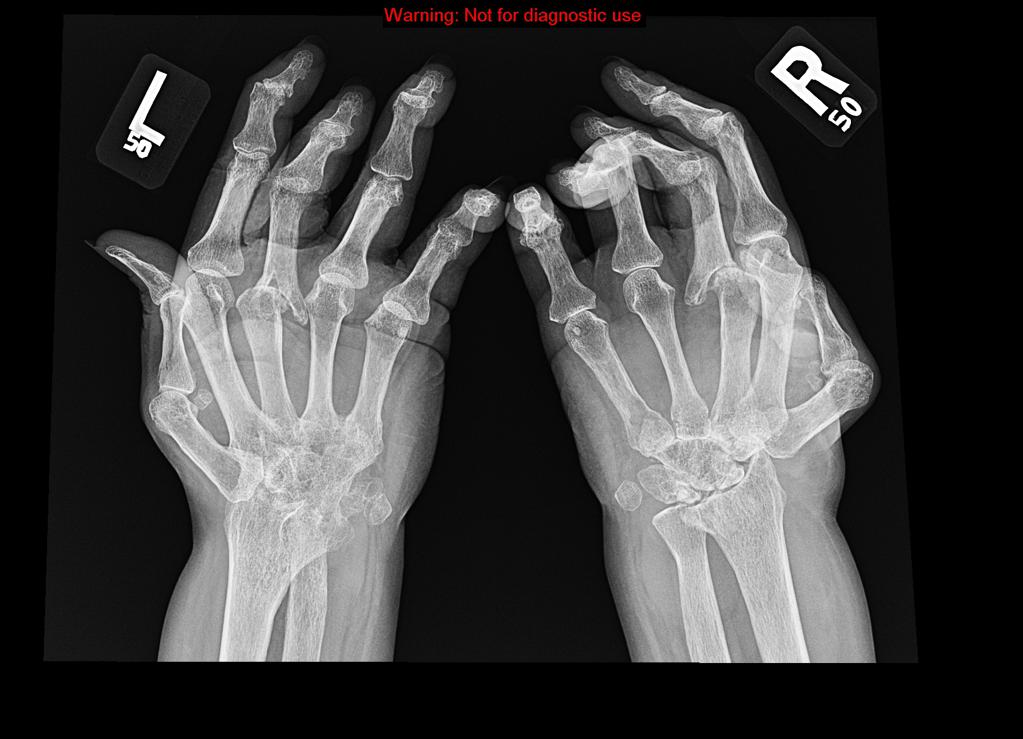
The findings may include:[2][3]
- Subchondral cysts
- Ulnar deviation of the MCP joints
- Boutonniere and swan neck deformities
- Hitchhiker’s thumb deformity
- Scapholunate dissociation, ulnar translocation
- Ankylosis

Source: A.Prof Frank Gaillard [4]

Source:Case courtesy of Dr Hani Salam[5]
Feet
Various radiological findings of the feet that may be seen in rheumatoid arthritis include:

Source: Case courtesy of Dr Ian Bickle, [6][7]
Shoulder
Various radiological findings of the shoulder joint that may be seen in rheumatoid arthritis include:
- Distal clavicle erosions
- Erosions of the superolateral aspect of the head of the humerus
- High riding shoulder due to subacromial-subdeltoid bursitis

Source:Case courtesy of Radswiki [8][9]
Knee
Various radiological findings of the knee joint that may be seen in rheumatoid arthritis include:[10]
- Joint effusions
- Loss of joint space
- Prepatellar bursitis
Hip
Various radiological findings of the hip joint that may be seen in rheumatoid arthritis include:
- Concentric loss of joint space
- Acetabular protrusio

Source:Case courtesy of Dr Jeremy Jones[11][12]
Spinal cord
Various radiological findings of the spinal cord joint that may be seen in rheumatoid arthritis include:
- Atlantoaxial subluxation
- Atlantoaxial impaction: cephalad migration of C2
- Osteoporosis and osteoporotic fractures
- Erosion of spinous processes

Source:Case courtesy of Dr Muhammad Essam, [13][14]
References
- ↑ Devauchelle-Pensec V, Saraux A, Alapetite S, Colin D, Le Goff P (October 2002). "Diagnostic value of radiographs of the hands and feet in early rheumatoid arthritis". Joint Bone Spine. 69 (5): 434–41. PMID 12477226.
- ↑ van der Heijde DM, van Leeuwen MA, van Riel PL, Koster AM, van 't Hof MA, van Rijswijk MH, van de Putte LB (January 1992). "Biannual radiographic assessments of hands and feet in a three-year prospective followup of patients with early rheumatoid arthritis". Arthritis Rheum. 35 (1): 26–34. PMID 1731813.
- ↑ Koh JH, Jung SM, Lee JJ, Kang KY, Kwok SK, Park SH, Ju JH (2015). "Radiographic Structural Damage Is Worse in the Dominant than the Non-Dominant Hand in Individuals with Early Rheumatoid Arthritis". PLoS ONE. 10 (8): e0135409. doi:10.1371/journal.pone.0135409. PMC 4527732. PMID 26247204.
- ↑ "https://radiopaedia.org, ref="https://radiopaedia.org/cases/7245">rID: 7245
- ↑ "https://radiopaedia.org/"Radiopaedia.org
- ↑ href="https://radiopaedia.org/">Radiopaedia.org
- ↑ href="https://radiopaedia.org/cases/27301">rID: 27301
- ↑ href="https://radiopaedia.org/">Radiopaedia.org
- ↑ href="https://radiopaedia.org/cases/11884">rID: 11884
- ↑ Fuchs HA, Kaye JJ, Callahan LF, Nance EP, Pincus T (May 1989). "Evidence of significant radiographic damage in rheumatoid arthritis within the first 2 years of disease". J. Rheumatol. 16 (5): 585–91. PMID 2754663.
- ↑ href="https://radiopaedia.org/">Radiopaedia.org
- ↑ href="https://radiopaedia.org/cases/6464">rID: 6464
- ↑ href="https://radiopaedia.org/">Radiopaedia.org
- ↑ href="https://radiopaedia.org/cases/29388">rID: 29388