|
|
| (8 intermediate revisions by 3 users not shown) |
| Line 1: |
Line 1: |
| | __NOTOC__ |
| {{SI}} | | {{SI}} |
| | | {{CMG}} |
| {{EH}} | |
|
| |
|
| ==Overview== | | ==Overview== |
| Line 8: |
Line 8: |
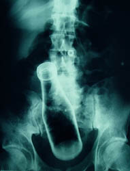
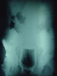
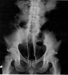
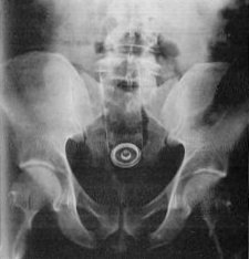
| Rectal foreign bodies, and amateur attempts to remove them, can result in [[perforation]] of the bowel, which is a life-threatening [[medical emergency]]. Medical literature covers examples of items retrieved from patients' rectums. Rectal foreign objects are also the subject of a number of urban legends. | | Rectal foreign bodies, and amateur attempts to remove them, can result in [[perforation]] of the bowel, which is a life-threatening [[medical emergency]]. Medical literature covers examples of items retrieved from patients' rectums. Rectal foreign objects are also the subject of a number of urban legends. |
|
| |
|
| ==Examples of foreign bodies== | | ==X Ray Examples== |
| | <gallery perRow="3"> |
|
| |
|
| <gallery perRow="3">
| |
| Image:Coin in esophagus 2.jpg|[[Chest X-ray]] showing a coin in the esophagus of a young child
| |
| Image:FB1.jpg| | | Image:FB1.jpg| |
| Image:FB2.jpg| | | Image:FB2.jpg| |
| Image:FB3.jpg| | | Image:FB3.jpg| |
| Image:screwdriver.jpg|A screwdriver with a plastic handle. (Dr. A.K. Sharma, Agra, India
| |
| Image:FB4.jpg| | | Image:FB4.jpg| |
| Image:Foreign Body.jpg|Gastric [[foreign body]] (toothbrush)
| |
| Image:FB6.JPG| | | Image:FB6.JPG| |
| Image:FB7.JPG| | | Image:FB7.JPG| |
| Image:FB8.jpg| | | Image:FB8.jpg| |
| Image:FB9.jpg|Gastric [[foreign body]] (toothbrush)
| |
| Image:Dildo03.jpg|Rectal foreign body (Image courtesy of Dr Frank Gaillard) | | Image:Dildo03.jpg|Rectal foreign body (Image courtesy of Dr Frank Gaillard) |
| Image:SlowK1.jpg|This patient collapsed on the ward and was thought to have had a pulmonary embolus. A CTPA revealed a tablet lying dependently in the patient's trachea, with changes of aspiration in both lower lobes (not shown). A second tablet was visible in the stomach... the tablet was removed via a bronchoscope and confirmed to be a Slow K (potassium). (Image courtesy of Dr Frank Gaillard)
| |
| Image:SlowK2.jpg|This patient collapsed on the ward and was thought to have had a pulmonary embolus. A CTPA revealed a tablet lying dependently in the patient's trachea, with changes of aspiration in both lower lobes (not shown). A second tablet was visible in the stomach... the tablet was removed via a bronchoscope and confirmed to be a Slow K (potassium). (Image courtesy of Dr Frank Gaillard)
| |
| Image:SlowK3.jpg|This patient collapsed on the ward and was thought to have had a pulmonary embolus. A CTPA revealed a tablet lying dependently in the patient's trachea, with changes of aspiration in both lower lobes (not shown). A second tablet was visible in the stomach... the tablet was removed via a bronchoscope and confirmed to be a Slow K (potassium). (Image courtesy of Dr Frank Gaillard)
| |
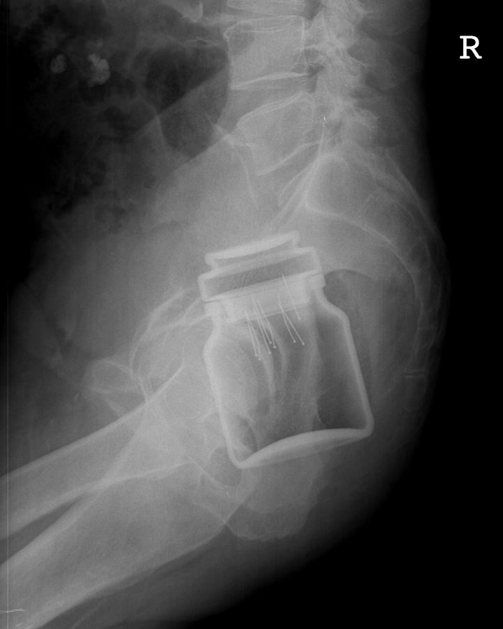
| Image:Jar722.jpg|Rectal foreign body (Image courtesy of Dr Frank Gaillard) | | Image:Jar722.jpg|Rectal foreign body (Image courtesy of Dr Frank Gaillard) |
| Image:Tampon_AXR.jpg|Vaginal foreign body (Image courtesy of Dr Donna D'Souza)
| |
| Image:Orbital_foreign_body.jpg|This patient presented with a self-harm injury. The axial CT scan shows a ball-point pen in-situ. The pen missed optic nerve, middle cerebral artery and any eloquent brain. A cerebral angiogram was performed which was normal except for truncation of the ophthalmic artery. The pen was removed under flouroscopic guidance. Upon removal, there was brisk bleeding from the ophthalmic artery. Endovascular embolisation of the bleeding vessel was performed with coils, with good result. The patient’s pupil remains reactive, suggesting a good prognosis for the optic nerve and the patient’s vision. (Image courtesy of Dr Laughlin Dawes)
| |
| Image:Mickey_mouse.JPG|A 7 year old girl presented to ED after swallowing a foreign body. Initial CXR shows a foreign body at the level of T8. There was no change in the position of the Mickey Mouse key ring after 8 hours. No pneumomediastinum or pneumothorax. The airway is patent. It was finally retrieved with oesophagoscopy. (Image courtesy of Dr Lily Wang)
| |
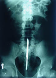
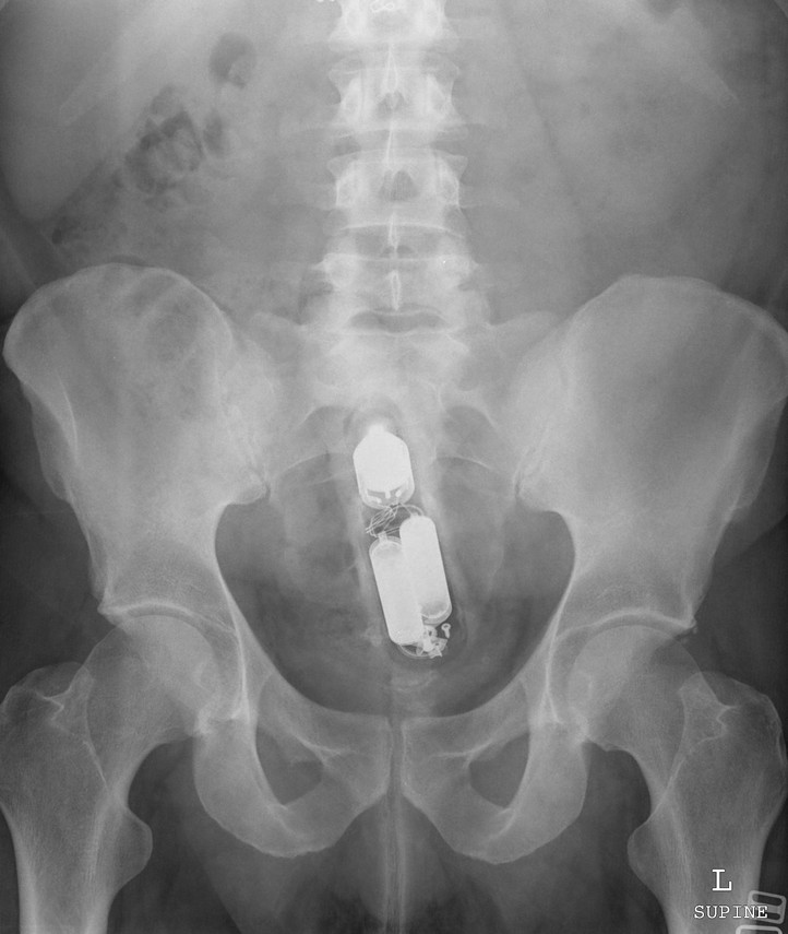
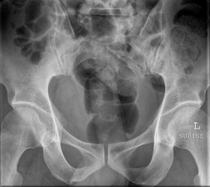
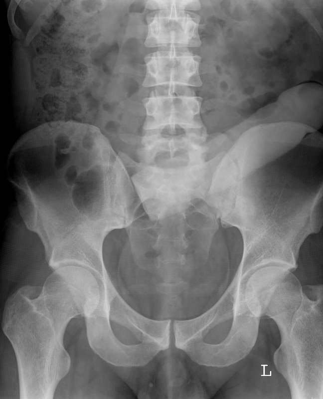
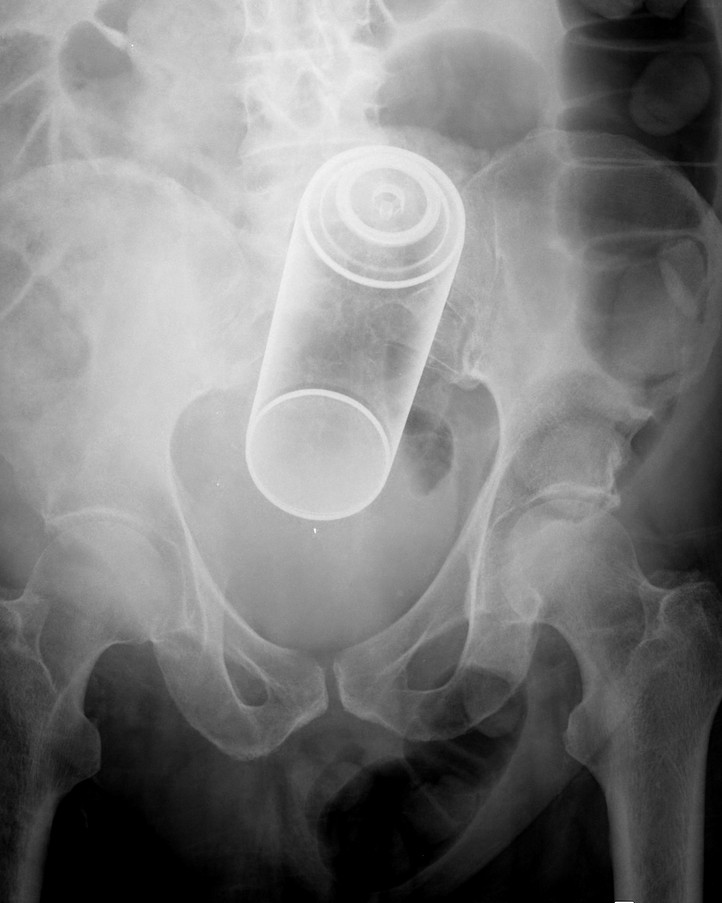
| Image:AXR_fb.JPG|A middle-aged male presented to the emergency department with abdominal discomfort. An abdominal radiograph was performed as shown. There is no evidence of perforation or obstruction. The patient was taken to operating room within 12 hours of presentation, with consent for colostomy. Under general anaesthesia in the lithotomy position, dilatation of anal sphincter was performed and per rectum retrieval successful. These patients typically have a delayed presentation to the emergency department because of embarrassment and after multiple attempts at self removal. Respect for their privacy is a key factor in the patient’s care plan. ED physicians need to decide if removal of foreign body can be performed in the emergency department or surgical team to be notified. Operating room procedures include anal dilatation under GA, transrectal manipulation, bimanual palpation if necessary and withdrawal of foreign body. Laparotomy or laparoscopy are occasionally necessary. (Image courtesy of Andrew Roshan)
| |
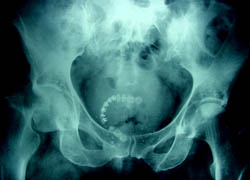
| Image:Body_Packer.jpg|Rectal foreign body. Body packer. (Image courtesy of Dr Frank Gaillard) | | Image:Body_Packer.jpg|Rectal foreign body. Body packer. (Image courtesy of Dr Frank Gaillard) |
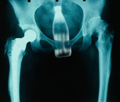
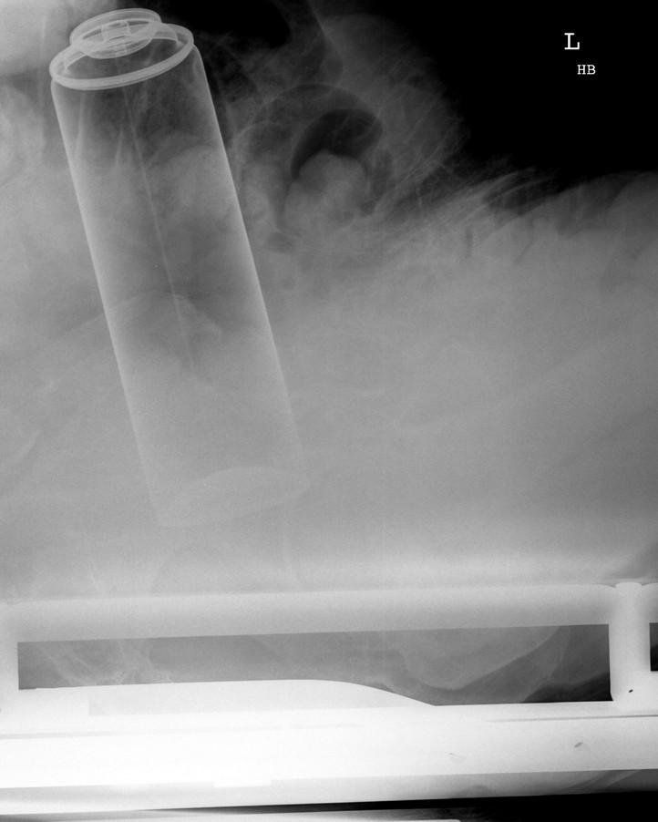
| Image:Sextoy.jpg|Rectal foreign body (Image courtesy of Dr Frank Gaillard) | | Image:Sextoy.jpg|Rectal foreign body (Image courtesy of Dr Frank Gaillard) |
| Image:RFB02.jpg|Rectal foreign body (Image courtesy of Dr Frank Gaillard) | | Image:RFB02.jpg|Rectal foreign body (Image courtesy of Dr Frank Gaillard) |
| Image:RFB03.jpg|Rectal foreign body (Image courtesy of Dr Frank Gaillard) | | Image:RFB03.jpg|Rectal foreign body (Image courtesy of Dr Frank Gaillard) |
| Image:Thong.JPG|(Image courtesy of Simon Pilgrim and Laughlin Dawes)
| |
| Image:Ring_pessary.jpg|Vaginal pessary for treatment of uterine prolapse (Image courtesy of Dr Frank Gaillard)
| |
| Image:Staple_gun_to_hand.jpg|This building-site worker inadvertently stapled-gunned his hand, neatly sitting in his interosseous space. (Image courtesy of Dr Frank Gaillard)
| |
| Image:Aspirated_tooth_CXR.jpg|This trauma patient had significant craniofacial injuries and aspirated a tooth. It lodged in the right lower lobe bronchus, causing post-obstructive consolidation. Inhaled foreign bodies may be complicated by haemoptysis, air trapping, post-obstructive collapse, pneumonia or bronchiectasis. Removal of the foreign body is usually performed via bronchoscopy. (Image courtesy of Dr Donna D'Souza)
| |
| Image:Wire_IVC01.jpg|This patient was transfered to ICU from another hospital. On examination of their abdomen a wire was noted to project through the right atrium, down the IVC to end in the right iliac vein. The J-shaped tip gave the game away... a right jugular CVC had been placed, and the wire used for the insertion pushed in with the catheter. This guide wire was successfully retrieved from the groin, without complication. (Image courtesy of Dr Frank Gaillard)
| |
| Image:Wire_IVC02.jpg|This patient was transfered to ICU from another hospital. On examination of their abdomen a wire was noted to project through the right atrium, down the IVC to end in the right iliac vein. The J-shaped tip gave the game away... a right jugular CVC had been placed, and the wire used for the insertion pushed in with the catheter. This guide wire was successfully retrieved from the groin, without complication. (Image courtesy of Dr Frank Gaillard)
| |
| </gallery> | | </gallery> |
|
| |
|
| == References in the media == | | ==Case Example== |
| | | [[Image:AXR_fb.JPG|center|200px]] |
| *The television show ''[[ER (tv series)|ER]]'' makes several references to this, such as when [[Peter Benton|Dr. Benton]] holds up a lower abdominal x-ray with a flashlight lodged in it, noting, "The patient claimed that he fell on it while changing a lightbulb. Naked." Another episode has a medical student asking [[John Carter (ER)|Dr. John Carter]] about the oddest object removed from a rectum. Dr. Carter responds that it was a bowling trophy.
| |
| | |
| *The television show ''[[Scrubs (tv series)|Scrubs]]'' has made two references to this phenomenon. In "[[My Two Dads (Scrubs)|My Two Dads]]" Turk gives his then girlfriend Carla a pen from what he thinks is the lost and found box, only to discover that the hospital has no lost and found box, and he has mistakenly given his girlfriend a pen from the "Ass Box," comprised entirely of items removed from peoples rectums. The second was when a patient came in with a lightbulb in his rectum, and Dr. Cox, Turk and the Janitor work together to figure out a way to remove the bulb without breaking it.
| |
| | |
| *The television show ''[[House (TV series)|House]]'' also made two references to rectal foreign bodies. In the first, [[Gregory House]] is tending to a clinic patient who refuses to sit, from which he deduces the object's size and location. Upon learning the patient has an MP3 player in his rectum, House asks "Is it because of the size, the shape ... or the pounding bass-line?". The second reference comes when House is confronting his boss, [[Lisa Cuddy]], explaining that the object he has won him second place in the "Weirdest Object Pulled From A Bodily Orifice" and that she does not want to know what the first place-getter was, but it does rhyme with "fucchini".
| |
| | |
| *On MTV's ''Jackass'', Ryan Dunn goes to the hospital with a [[Hot Wheels|HotWheel]] wrapped in a [[condom]] in his rectum.
| |
| | |
| *On an [[The Fusilli Jerry|episode]] of [[Seinfeld]], [[George Costanza]]'s father falls on a small sculpture that [[Cosmo Kramer]] made, resulting in it being lodged in Mr. Costanza's rectum.
| |
|
| |
|
| == References ==
| | A middle-aged male presented to the emergency department with [[abdominal discomfort]]. An abdominal radiograph was performed as shown. There is no evidence of perforation or [[obstruction]]. The patient was taken to operating room within 12 hours of presentation, with consent for [[colostomy]]. Under [[general anesthesia]] in the [[lithotomy]] position, dilatation of [[anal sphincter]] was performed and per rectum retrieval successful. These patients typically have a delayed presentation to the emergency department because of embarrassment and after multiple attempts at self removal. Respect for their privacy is a key factor in the patient’s care plan. ED physicians need to decide if removal of foreign body can be performed in the emergency department or surgical team to be notified. Operating room procedures include anal dilatation under GA, transrectal manipulation, bimanual palpation if necessary and withdrawal of foreign body. [[Laparotomy]] or [[laparoscopy]] are occasionally necessary. (Image courtesy of Andrew Roshan) |
| * Busch D B, Starling J R. Rectal foreign bodies: Case reports and a comprehensive review of the world's literature. ''Surg'' 1986; '''100''': 512-519. See also the [[List of Ig Nobel Prize winners#1995|1995 Literature laureates]] of the [[Ig Nobel Prize]].
| |
|
| |
|
| == External links == | | ==References== |
| * Cynsa's [http://www.well.com/user/cynsa/newbutt.html Rectal Foreign Bodies page]
| | {{Reflist|2}} |
| * [http://www.emedicine.com/emerg/topic933.htm eMedicine gastrointestinal emergency medicine]
| |
| * [http://www.mja.com.au/public/issues/xmas98/mkinnon/mkinnon.html Medical Journal of Australia]
| |
| * [http://www.edu.rcsed.ac.uk/Case%20Presentations/CP22.htm Royal College of Surgeons of Edinburgh]
| |
| * British Dental Journal case report: [http://www.nature.com/cgi-taf/DynaPage.taf?file=/bdj/journal/v191/n1/full/4801082a.html Don't forget your toothbrush!] (subscribers only - with useful bibliography)
| |
|
| |
|
| [[Category:Emergency medicine]] | | [[Category:Emergency medicine]] |
| [[Category:Anal eroticism]]
| |
| [[nl:Rectal foreign object]]
| |
|
| |
|
| {{WH}} | | {{WH}} |
| {{WS}} | | {{WS}} |