Pyelonephritis CT scan: Difference between revisions
No edit summary |
|||
| Line 1: | Line 1: | ||
__NOTOC__ | __NOTOC__ | ||
{{Pyelonephritis}} | {{Pyelonephritis}} | ||
{{CMG}} | {{CMG}} | ||
==Overview== | ==Overview== | ||
==CT== | ==CT== | ||
===Acute Pyelonephritis=== | ===Acute Pyelonephritis=== | ||
[http://www.radswiki.net Images courtesy of RadsWiki] | [http://www.radswiki.net Images courtesy of RadsWiki] | ||
<gallery> | <gallery> | ||
Image:Acute pyelonephritis 001.jpg|CT: Acute pyelonephritis | Image:Acute pyelonephritis 001.jpg|CT: Acute pyelonephritis | ||
| Line 19: | Line 12: | ||
Image:Acute pyelonephritis 003.jpg|CT: Acute pyelonephritis | Image:Acute pyelonephritis 003.jpg|CT: Acute pyelonephritis | ||
</gallery> | </gallery> | ||
===Chronic Pyelonephritis=== | ===Chronic Pyelonephritis=== | ||
Imaging findings are characterized by renal scarring, [[atrophy]] and cortical thinning, [[hypertrophy]] of residual normal tissue, caliceal clubbing secondary to retraction of the [[papilla]] from overlying scar, thickening and dilatation of the caliceal system, and overall renal asymmetry. | |||
Imaging findings are characterized by renal scarring, atrophy and cortical thinning, hypertrophy of residual normal tissue, caliceal clubbing secondary to retraction of the papilla from overlying scar, thickening and dilatation of the caliceal system, and overall renal asymmetry. | |||
[http://www.radswiki.net Images courtesy of RadsWiki] | [http://www.radswiki.net Images courtesy of RadsWiki] | ||
<gallery> | <gallery> | ||
Image:Chronic-pyelonephritis-001.jpg|CT image demonstrates chronic pyelonephritis on the right | Image:Chronic-pyelonephritis-001.jpg|CT image demonstrates chronic pyelonephritis on the right | ||
| Line 31: | Line 20: | ||
Image:Chronic-pyelonephritis-003.jpg|CT image demonstrates chronic pyelonephritis on the right | Image:Chronic-pyelonephritis-003.jpg|CT image demonstrates chronic pyelonephritis on the right | ||
</gallery> | </gallery> | ||
===Emphysematous Pyelonephritis=== | ===Emphysematous Pyelonephritis=== | ||
*Additional evaluation with CT will confirm the presence and extent of parenchymal gas and will often allow identification of the source of obstruction when present. | *Additional evaluation with CT will confirm the presence and extent of parenchymal gas and will often allow identification of the source of obstruction when present. | ||
*The use of intravenous contrast material will often reveal asymmetric renal enhancement or delayed excretion, and, during the nephrographic phase, will help identify areas of focal tissue necrosis or abscess formation. | *The use of intravenous contrast material will often reveal asymmetric renal enhancement or delayed excretion, and, during the [[nephrographic]] phase, will help identify areas of focal tissue [[necrosis]] or abscess formation. | ||
[http://www.radswiki.net Images courtesy of RadsWiki] | [http://www.radswiki.net Images courtesy of RadsWiki] | ||
<gallery> | <gallery> | ||
Image:Emphysematous-pyelonephritis-001.jpg|CT: Emphysematous pyelonephritis | Image:Emphysematous-pyelonephritis-001.jpg|CT: Emphysematous pyelonephritis | ||
| Line 44: | Line 29: | ||
Image:Emphysematous-pyelonephritis-003.jpg|CT: Emphysematous pyelonephritis | Image:Emphysematous-pyelonephritis-003.jpg|CT: Emphysematous pyelonephritis | ||
</gallery> | </gallery> | ||
===Xanthogranulomatous Pyelonephritis=== | ===Xanthogranulomatous Pyelonephritis=== | ||
The CT findings of xanthogranulomatous pyelonephritis are pathognomonic in most cases: diffuse [[reniform]] enlargement with ill-defined central low attenuation, apparent cortical thinning, and central calculi. | |||
The CT findings of xanthogranulomatous pyelonephritis are pathognomonic in most cases: diffuse reniform enlargement with ill-defined central low attenuation, apparent cortical thinning, and central calculi. | **Extension into the [[perinephric space]] and beyond the [[Gerota fascia]] is not uncommon. | ||
**Extension into the perinephric space and beyond the Gerota fascia is not uncommon. | **Central areas of low [[attenuation]] represent [[nonenhancing]] xanthomatous material that may demonstrate attenuation values less than those of water. | ||
**Central areas of low attenuation represent nonenhancing xanthomatous material that may demonstrate attenuation values less than those of water. | |||
[http://www.radswiki.net Images courtesy of RadsWiki] | [http://www.radswiki.net Images courtesy of RadsWiki] | ||
<gallery> | <gallery> | ||
Image:Xanthogranulomatous pyelonephritis 001.jpg|CT image demonstrates right xanthogranulomatous pyelonephritis | Image:Xanthogranulomatous pyelonephritis 001.jpg|CT image demonstrates right xanthogranulomatous pyelonephritis | ||
| Line 63: | Line 44: | ||
[[Category:Needs content]] | [[Category:Needs content]] | ||
[[Category:Nephrology]] | |||
[[Category:Inflammations]] | |||
[[Category:Disease]] | |||
{{WH}} | {{WH}} | ||
{{WS}} | {{WS}} | ||
Revision as of 15:00, 27 September 2012
|
Pyelonephritis Microchapters |
|
Diagnosis |
|
Treatment |
|
Case Studies |
|
Pyelonephritis CT scan On the Web |
|
American Roentgen Ray Society Images of Pyelonephritis CT scan |
|
Risk calculators and risk factors for Pyelonephritis CT scan |
Editor-In-Chief: C. Michael Gibson, M.S., M.D. [1]
Overview
CT
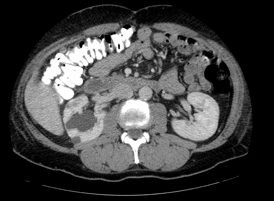
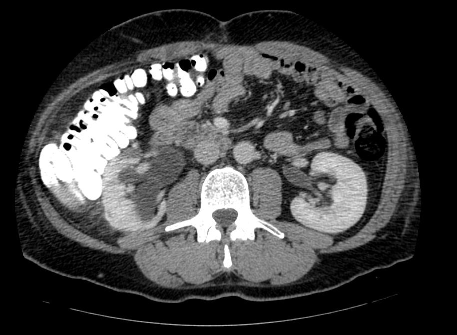
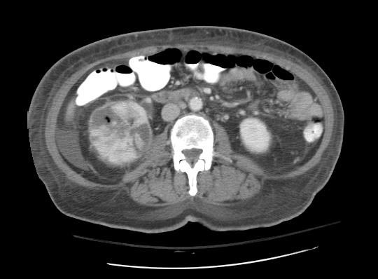
Acute Pyelonephritis
-
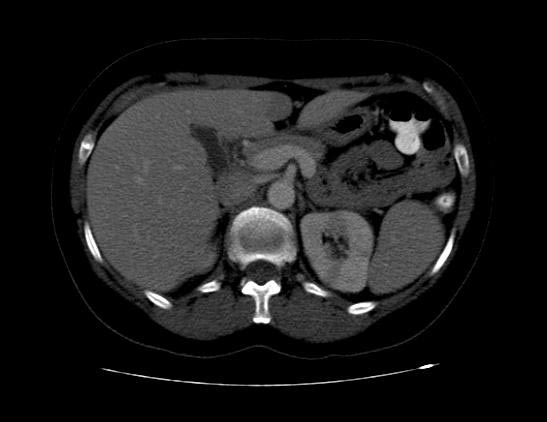
CT: Acute pyelonephritis
-
CT: Acute pyelonephritis
-
CT: Acute pyelonephritis


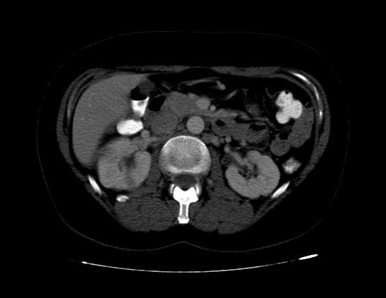
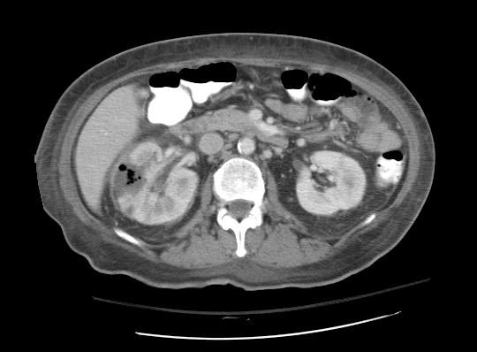
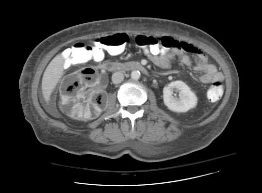
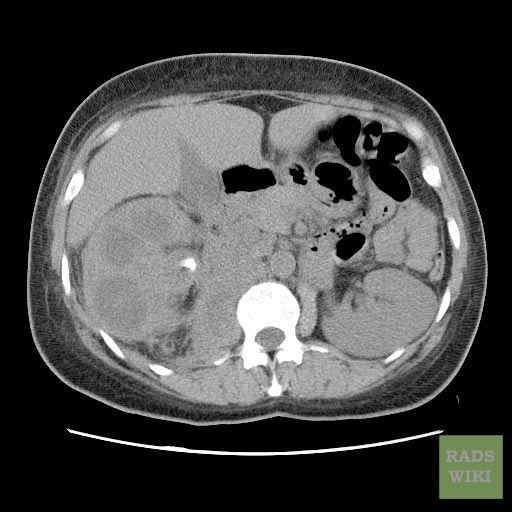
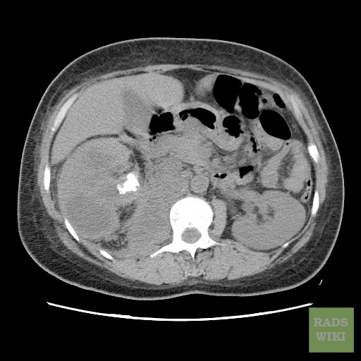
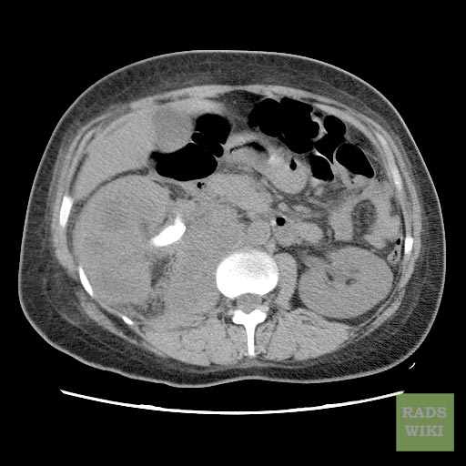
Chronic Pyelonephritis
Imaging findings are characterized by renal scarring, atrophy and cortical thinning, hypertrophy of residual normal tissue, caliceal clubbing secondary to retraction of the papilla from overlying scar, thickening and dilatation of the caliceal system, and overall renal asymmetry. Images courtesy of RadsWiki
-
CT image demonstrates chronic pyelonephritis on the right
-
CT image demonstrates chronic pyelonephritis on the right
-
CT image demonstrates chronic pyelonephritis on the right


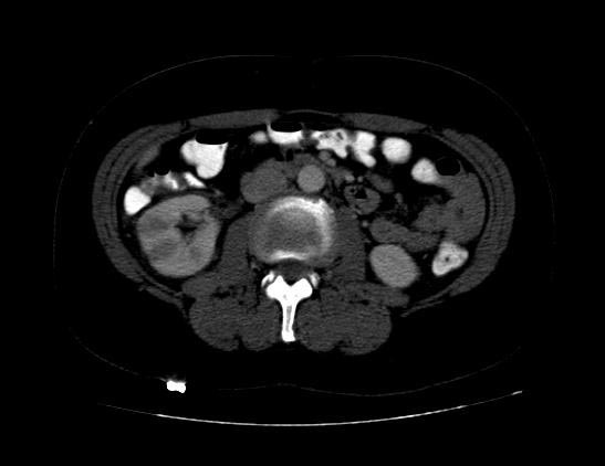
Emphysematous Pyelonephritis
- Additional evaluation with CT will confirm the presence and extent of parenchymal gas and will often allow identification of the source of obstruction when present.
- The use of intravenous contrast material will often reveal asymmetric renal enhancement or delayed excretion, and, during the nephrographic phase, will help identify areas of focal tissue necrosis or abscess formation.
-
CT: Emphysematous pyelonephritis
-
CT: Emphysematous pyelonephritis
-
CT: Emphysematous pyelonephritis


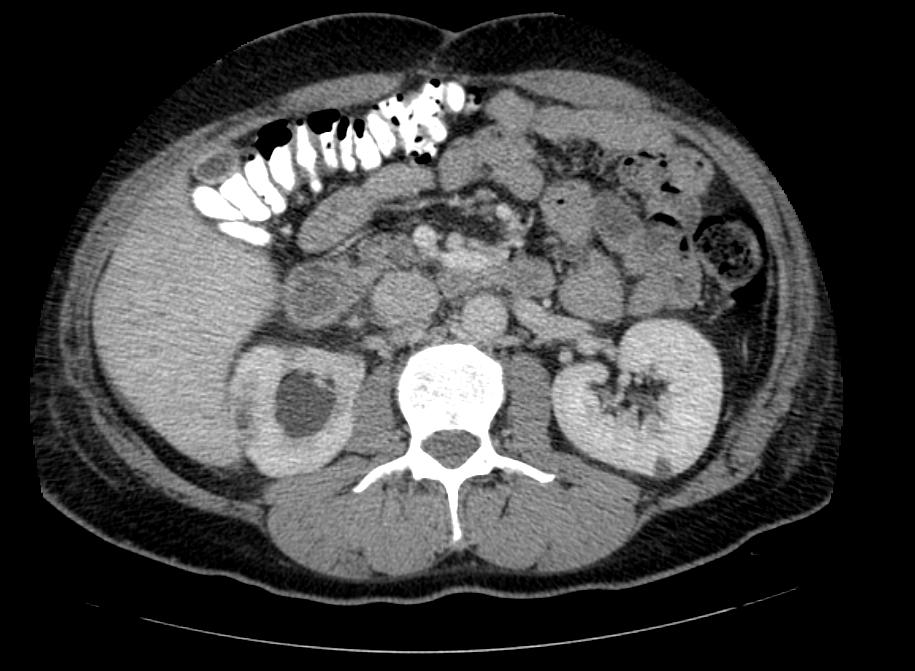
Xanthogranulomatous Pyelonephritis
The CT findings of xanthogranulomatous pyelonephritis are pathognomonic in most cases: diffuse reniform enlargement with ill-defined central low attenuation, apparent cortical thinning, and central calculi.
- Extension into the perinephric space and beyond the Gerota fascia is not uncommon.
- Central areas of low attenuation represent nonenhancing xanthomatous material that may demonstrate attenuation values less than those of water.
-
CT image demonstrates right xanthogranulomatous pyelonephritis
-
CT image demonstrates right xanthogranulomatous pyelonephritis
-
CT image demonstrates right xanthogranulomatous pyelonephritis


