Pulmonary nodule CT: Difference between revisions
No edit summary |
|||
| Line 83: | Line 83: | ||
| Follow-up at 12 months. If no change, no further imaging needed | | Follow-up at 12 months. If no change, no further imaging needed | ||
|- | |- | ||
| >4 - 6 | | > 4 - 6 | ||
| Follow-up at 12 months. If no change, no further imaging needed | | Follow-up at 12 months. If no change, no further imaging needed | ||
| Initial follow-up CT at 6 -12 months and then at 18 - 24 months if no change | | Initial follow-up CT at 6 -12 months and then at 18 - 24 months if no change | ||
|- | |- | ||
| >6 - 8 | | > 6 - 8 | ||
| Initial follow-up CT at 6 -12 months and then at 18 - 24 months if no change | | Initial follow-up CT at 6 -12 months and then at 18 - 24 months if no change | ||
| Initial follow-up CT at 3 - 6 months and then at 9 -12 and 24 months if no change | | Initial follow-up CT at 3 - 6 months and then at 9 -12 and 24 months if no change | ||
|- | |- | ||
| >8 | | > 8 | ||
| Follow-up CTs at around 3, 9, and 24 months. Dynamic contrast enhanced CT, PET, and/or biopsy | | Follow-up CTs at around 3, 9, and 24 months. Dynamic contrast enhanced CT, PET, and/or biopsy | ||
| Same at for low risk patients | | Same at for low risk patients | ||
Revision as of 14:05, 19 March 2016
|
Pulmonary Nodule Microchapters |
|
Diagnosis |
|---|
|
Treatment |
|
Case Studies |
|
Pulmonary nodule CT On the Web |
|
American Roentgen Ray Society Images of Pulmonary nodule CT |
Editor-In-Chief: C. Michael Gibson, M.S., M.D. [1]Associate Editor(s)-in-Chief: Maria Fernanda Villarreal, M.D. [2]
Overview
Computed tomography is the method of choice for the diagnosis of solitary pulmonary nodule. On CT, characteristic findings of solitary pulmonary nodules, include: ground-glass opacity, rounded mass, and less than 30mm.[1][2] The evaluation of solitary pulmonary nodule will depend on 7 characteristics: calcification patterns, size, location, size, growth, shape, margins, attenuation, and contrast enhancement.[2]
CT
- Computed tomography is the method of choice for the diagnosis of solitary pulmonary nodule
- On CT, characteristic findings of solitary pulmonary nodules, include:
- Single intraparenchymal lesion
- Less than 3 cm in size
- Rounded or spicluated lesion
The evaluation of solitary pulmonary nodule will depend on the following characteristics:
Calcification
- Calcification patterns are commonly seen in granulomatous disease and hamartomas
- Characteristic calcification patterns of pulmonary nodule, include:
- Diffuse
- Central
- Laminated
- Popcorn
Size
- Different size ranges of pulmonary nodule, include:
- Nodules less than 4mm
- Nodules between 4mm and 7mm
- Nodules between 8mm and 20mm
- Nodules more than 20mm
Growth
- The growth pattern of the pulmonary nodule plays an important role in the management strategy.[3]
- Nodule growth should be evaluated on a individual basis and based on the risk assessment score
- A 4x growth is associated with a 50% risk of malignancy[3]
Shape
- Polygonal
- Spherical
Margins
- Lobulated or scalloped margins
- Intermediate malignancy probability
- Smooth margins
- Associated with nodule benignancy
Attenuation
- Different types of attenuation for pulmonary nodule, include:
- Solid pulmonary nodules
- Malignancy rate of only 7%
- Calcified pulmonary nodules
- Partly solid pulmonary nodules
- Malignancy rate of 63%
- Ground glass pulmonary nodules
- Malignancy rate of 18%
Contrast enhancement
- Contrast enhancement of pulmonary nodules may be useful to determine benign or malignant features
- Benign pulmonary nodules usually have a contrast enhancement less than 15 HU
On CT, radiological signs of pulmonary nodule, include:
- Corona radiata sign: highly associated with malignancy
- Air bronchogram sign: airway surrounded by collection in alveolar spaces, non-specific sign
- Halo sign: zone of ground-glass attenuation surrounding a pulmonary nodule or mass on CT images
CT Surveillance
According to the American College of Chest Physicians (ACCP) for the CT surveillance of pulmonary nodules, recommends the following:[4]
- If less than 8 mm, use guidelines by the Fleischner society (see table below).
- For nodules greater than 8 mm in diameter, assess the patients risk of complications from thoracic surgery:
- If low to moderate risk for complications of surgery, assess probability of cancer by a validated calculation. The model developed at the Mayo Clinic has been the most extensively validated. An open-source version is available online.
- If high risk for complications of surgery, assess probability of cancer by a validated calculation. If low to moderate risk of cancer follow up with CT scan surveillance. If moderate to high risk of cancer obtain non-surgical biopsy.[5]
| Nodule Size (mm) | Low risk patients† | High risk patients‡ |
|---|---|---|
| <= 4 | No follow-up needed | Follow-up at 12 months. If no change, no further imaging needed |
| > 4 - 6 | Follow-up at 12 months. If no change, no further imaging needed | Initial follow-up CT at 6 -12 months and then at 18 - 24 months if no change |
| > 6 - 8 | Initial follow-up CT at 6 -12 months and then at 18 - 24 months if no change | Initial follow-up CT at 3 - 6 months and then at 9 -12 and 24 months if no change |
| > 8 | Follow-up CTs at around 3, 9, and 24 months. Dynamic contrast enhanced CT, PET, and/or biopsy | Same at for low risk patients |
| † Low risk patients: Minimal or absent history of smoking and of other known risk factors. ‡ High risk patients: History of smoking or of other known risk factors | ||
Gallery
-

Halo sign: Ground glass opacity surrounding a pulmonary nodule
-

Thorax CT
-
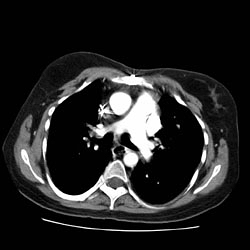
Thorax CT


References
- ↑ Rosado-de-Christenson ML, Templeton PA, Moran CA (1994). "Bronchogenic carcinoma: radiologic-pathologic correlation". Radiographics. 14 (2): 429–46, quiz 447–8. doi:10.1148/radiographics.14.2.8190965. PMID 8190965.
- ↑ 2.0 2.1 Parker MS, Chasen MH, Paul N (2009). "Radiologic signs in thoracic imaging: case-based review and self-assessment module". AJR Am J Roentgenol. 192 (3 Suppl): S34–48. doi:10.2214/AJR.07.7081. PMID 19234288.
- ↑ 3.0 3.1 Ko JP, Berman EJ, Kaur M, Babb JS, Bomsztyk E, Greenberg AK, Naidich DP, Rusinek H (2012). "Pulmonary Nodules: growth rate assessment in patients by using serial CT and three-dimensional volumetry". Radiology. 262 (2): 662–71. doi:10.1148/radiol.11100878. PMC 3267080. PMID 22156993.
- ↑ Gould MK, Donington J, Lynch WR, Mazzone PJ, Midthun DE, Naidich DP; et al. (2013). "Evaluation of individuals with pulmonary nodules: when is it lung cancer? Diagnosis and management of lung cancer, 3rd ed: American College of Chest Physicians evidence-based clinical practice guidelines". Chest. 143 (5 Suppl): e93S–120S. doi:10.1378/chest.12-2351. PMC 3749714. PMID 23649456.
- ↑ Swensen SJ, Silverstein MD, Ilstrup DM, Schleck CD, Edell ES (1997). "The probability of malignancy in solitary pulmonary nodules. Application to small radiologically indeterminate nodules". Arch Intern Med. 157 (8): 849–55. PMID 9129544.
- ↑ MacMahon H, Austin JH, Gamsu G, Herold CJ, Jett JR, Naidich DP; et al. (2005). "Guidelines for management of small pulmonary nodules detected on CT scans: a statement from the Fleischner Society". Radiology. 237 (2): 395–400. doi:10.1148/radiol.2372041887. PMID 16244247.