Pulmonary hypertension chest x ray
|
Pulmonary Hypertension Microchapters |
|
Diagnosis |
|---|
|
Treatment |
|
Case Studies |
|
Pulmonary hypertension chest x ray On the Web |
|
American Roentgen Ray Society Images of Pulmonary hypertension chest x ray |
|
Risk calculators and risk factors for Pulmonary hypertension chest x ray |
Editor-In-Chief: C. Michael Gibson, M.S., M.D. [1], Richard Channick, M.D.; Assistant Editor(s)-in-Chief: Ralph Matar; José Eduardo Riceto Loyola Junior, M.D.[2]
Overview
A chest X-ray is abnormal in the majority of patients with pulmonary hypertension (PH); however, there is no correlation between the severity of PH and the findings on a chest X-ray. Findings of PH on a chest X-ray include pulmonary artery dilatation and right-sided enlargement of the heart. A chest X-ray may suggest that there is no compromise of the left heart if normal and allows for initial assessment of lung disease that can lead to group 2 and group 3 PH, respectively.
Chest X Ray
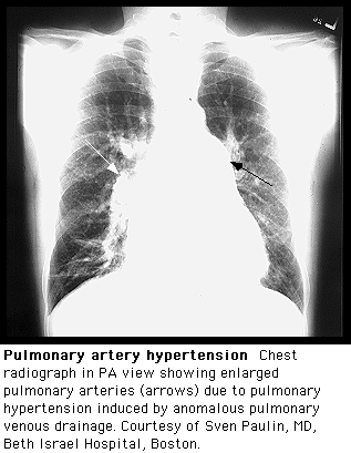
Findings of PH on a chest X-ray include:[1][2][3]
- Hilar pulmonary arterial dilation
- Loss of peripheral blood vessel markings
- Enlarged right atrium, right ventricle and pulmonary arteries in advanced diseases.[4]
Shown below are chest X-ray images of patients with PH.
-
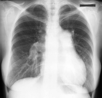
This is a posteroanterior radiograph revealing enlarged pulmonary arteries in a patient with atrial septal defect.
-


References
- ↑ Korobkova IZ, Lazutkina VK, Nizovtsova LA, Riden TV (2015). "[Radiographic assessment of pulmonary hypertension: Methodical aspects]". Vestn Rentgenol Radiol (in Russian) (4): 45–53. PMID 26552229.
- ↑ Pienn M, Kovacs G, Tscherner M, Avian A, Johnson TR, Kullnig P, Stollberger R, Olschewski A, Olschewski H, Bálint Z (March 2014). "Non-invasive determination of pulmonary hypertension with dynamic contrast-enhanced computed tomography: a pilot study". Eur Radiol. 24 (3): 668–76. doi:10.1007/s00330-013-3067-8. PMID 24311231.
- ↑ Cordova FC, D'Alonzo G (September 2013). "Sarcoidosis-associated pulmonary hypertension". Curr Opin Pulm Med. 19 (5): 531–7. doi:10.1097/MCP.0b013e328363f4a3. PMID 23912192.
- ↑ Poch D, Mandel J (2021). "Pulmonary Hypertension". Ann Intern Med. 174 (4): ITC49–ITC64. doi:10.7326/AITC202104200. PMID 33844574 Check
|pmid=value (help).