Pulmonary atresia (patient information): Difference between revisions
No edit summary |
|||
| Line 43: | Line 43: | ||
:*[[Echocardiography]]: This kind of painless test can help the doctor closely examine pulmonary atresia. It uses sound waves to produce an image of the ventricles, atrium and great vessles. Echocardiogram can tell whether pulmonary valve opens to let blood flow through or not. Further more, the doctor can measure the speed of blood flow through patient's heart and pulmonary valve area by echocardiogram. | :*[[Echocardiography]]: This kind of painless test can help the doctor closely examine pulmonary atresia. It uses sound waves to produce an image of the ventricles, atrium and great vessles. Echocardiogram can tell whether pulmonary valve opens to let blood flow through or not. Further more, the doctor can measure the speed of blood flow through patient's heart and pulmonary valve area by echocardiogram. | ||
:*[[Chest x-ray]]: An x-ray image of chest allows the doctor to check the size and shape of your heart to determine whether the left atrium and ventricle are enlarged or not. And it also helps the doctor check the condition of your lungs. Patients with pulmonary atresia may show enlarged right ventricle on an x -ray. | :*[[Chest x-ray]]: An x-ray image of chest allows the doctor to check the size and shape of your heart to determine whether the left atrium and ventricle are enlarged or not. And it also helps the doctor check the condition of your lungs. Patients with pulmonary atresia may show enlarged right ventricle on an x -ray. | ||
:*Chest CT or MRI: A chest CT or MRI can demonstrate the details of the heart extremely well, such as the positions of valval, vascular, atrial and ventricular structures and their relationships to one another. | :*Chest CT or [[MRI]]: A chest CT or MRI can demonstrate the details of the heart extremely well, such as the positions of valval, vascular, atrial and ventricular structures and their relationships to one another. | ||
:*[[Electrocardiogram]] (ECG) and [[Holter monitoring]]: Electrocardiogram and Holter monitoring | :*[[Electrocardiogram]] (ECG) and [[Holter monitoring]]: Electrocardiogram and Holter monitoring detect electric activities of the heart for cardiovascular diseases. They can supply information about heart rhythm and indirectly, heart size. Patients with patent ductus pulmonary atresia may show a enlarged right ventricle sign. | ||
==When to seek urgent medical care?== | ==When to seek urgent medical care?== | ||
Revision as of 20:32, 18 February 2010
For the WikiDoc page for this topic, click here
| Pulmonary atresia | ||
 | ||
|---|---|---|
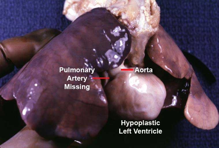
| Only an aorta can be seen originating from this pathology specimen. No pulmonary artery is present. | ||
| ICD-10 | Q25.5 | |
| ICD-9 | 747.3 | |
| MedlinePlus | 001091 | |
| eMedicine | ped/2526 ped/2898 | |
| MeSH | C14.240.670 | |
Editor-In-Chief: C. Michael Gibson, M.S., M.D. [1]; Jinhui Wu, MD
Please Join in Editing This Page and Apply to be an Editor-In-Chief for this topic: There can be one or more than one Editor-In-Chief. You may also apply to be an Associate Editor-In-Chief of one of the subtopics below. Please mail us [2] to indicate your interest in serving either as an Editor-In-Chief of the entire topic or as an Associate Editor-In-Chief for a subtopic. Please be sure to attach your CV and or biographical sketch.
What is pulmonary atresia?
Pulmonary atresia is an extremely rare form of congenital heart disease in which the pulmonary valve does not form properly. This leads blood from the right side of the heart cannot go to the lungs to pick up oxygen. Also, the right ventricle and tricuspid valve are often poorly developed. Pulmonary atresia is always associated with other congenital heart disease such as ventricular septal defect, tetralogy of Fallot and patent ductus arteriosus. The cause is not clear. Usual symptoms are cyanosis, shortness of breath, fast breathing and fatigue. Echocardiography and cardiac MRI can tell the diagnosis of pulmonary atresia. Treatments include prostaglandin E1, catheter-based procedures and surgery. Prognosis of pulmonary atresia depends on whether the surgery has been done and the severity of other associated congenital heart diseases.
How do I know if I have pulmonary atresia and what are the symptoms of pulmonary atresia?
Symptoms usually occur in the first few hours of life. Symptoms may include:
- Cyanosis
- Shortness of breath
- Fast breathing
- Poor eating habits
- Fatigue
Other health problems may also cause these symptoms. Only a doctor can tell for sure. A person with any of these symptoms should tell the doctor so that the problems can be diagnosed and treated as early as possible.
Who is at risk for pulmonary atresia?
Like other congenital heart diseases, the cause of pulmonary atresia is not clear. Pulmonary atresia sometimes is associated with other congenital heare diseases such as patent ductus arteriosus, ventricular septal defect and tetralogy of Fallot.
How to know you have pulmonary atresia?
- Echocardiography: This kind of painless test can help the doctor closely examine pulmonary atresia. It uses sound waves to produce an image of the ventricles, atrium and great vessles. Echocardiogram can tell whether pulmonary valve opens to let blood flow through or not. Further more, the doctor can measure the speed of blood flow through patient's heart and pulmonary valve area by echocardiogram.
- Chest x-ray: An x-ray image of chest allows the doctor to check the size and shape of your heart to determine whether the left atrium and ventricle are enlarged or not. And it also helps the doctor check the condition of your lungs. Patients with pulmonary atresia may show enlarged right ventricle on an x -ray.
- Chest CT or MRI: A chest CT or MRI can demonstrate the details of the heart extremely well, such as the positions of valval, vascular, atrial and ventricular structures and their relationships to one another.
- Electrocardiogram (ECG) and Holter monitoring: Electrocardiogram and Holter monitoring detect electric activities of the heart for cardiovascular diseases. They can supply information about heart rhythm and indirectly, heart size. Patients with patent ductus pulmonary atresia may show a enlarged right ventricle sign.
When to seek urgent medical care?
Call your health care provider if your baby has symptoms of pulmonary atresia. If one emerges the following symptoms, seeking urgent medical care as soon as possible:
Treatment options
Patients with pulmonart atresia have many treatment options. The selection depends on the size of the pulmonary artery and right ventricle, other associated heart defect and whether the child occurs the complications or not. The options are medicines, catheter-based procedures and surgery. Talk to your child's doctor about treatment options and your family's preferences on treatment decisions.
- Medicines: Prostaglandin E1 is usually used to help the blood move into the lungs.
- Catheter-based procedures: In a catheter room, the doctor threads a thin tube through a blood vessel in your baby's arm or groin to an artery in the heart and injects dye to see the heart and the arteries on an x-ray. Then a small metal coil or other blocking device is passed up through the catheter and placed to repair the defects. Complications of catheter-based procedures may include bleeding, infection and arrhythmia.
- Surgery: Surgical treatment of pulmonary atresia may be performed to repair or replace the valve. This surgery can also be used to place a tube between the right ventricle and the pulmonary arteries. During the surgery, the anesthetist gives medicine to make the child sleepy and comfortable. Then the surgeons make a small cut between the ribs to reach the heart and repair the defects and place a tube between the right ventricular and the plmonary arteries. Most cases can be helped with surgery.
Diseases with similar symptoms
Where to find medical care for pulmonary atresia?
Directions to Hospitals Treating pulmonary atresia
Prevention of pulmonary atresia
Preventive measure is unknown. Many congenital defects can be discovered on routine ultrasound examinations. All pregnant women should receive good prenatal care. Screening test and regular check for those with family history of congenital heart disease may be helpful.
What to expect (Outook/Prognosis)?
Most cases can be helped with surgery. Prognosis of pulmonary atresia depends on:
- Whether the corrective surgery has been done or not.
- The seveity of other associated congenital heart diseases.
Copyleft Sources
http://www.nlm.nih.gov/medlineplus/ency/article/001091.htm
http://www.americanheart.org/presenter.jhtml?identifier=1303